
In 2016, the pioneer of medical diagnostic imaging Toshiba Medical Systems Corporation (now Canon Medical Systems Corporation) joined the Canon Group. The synergy between Canon Medical’s diagnostic imaging systems and Canon’s optical and image processing technologies positions the Canon Group at the forefront of efforts to build a better future for a society.
2018/12/27Featured Technology
The history of Canon’s medical device business reaches as far back as 1940, shortly after its foundation, when the company developed Japan’s first indirect X-ray camera. Since then, Canon has leveraged the optical and image-processing technologies cultivated through camera development to introduce such leading-edge medical equipment as digital radiography systems and ophthalmic diagnostic equipment. In order to take its medical business to the next level, Canon welcomed Toshiba Medical Systems Corporation (now Canon Medical Systems Corporation) into the Canon Group in December 2016. Canon Medical boasts the top share of Computed Tomography (CT) systems in the Japanese market and ranks third* globally.
Since its development, CT technology has continued to evolve in response to demand from the medical community for imaging that is wider, faster and realizes higher resolution, all while reducing patient radiation doses.
Wider imaging was achieved with the introduction of the Aquilion ONE in 2007, which featured a 320-row area detector capable of scanning a 16 cm-wide section of the body with each rotation. The faster system also made possible the capture of image data for such major internal organs as the heart or brain with only a single rotation at a maximum scanning speed of 0.275 seconds per rotation.
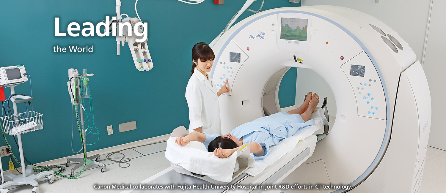
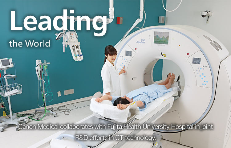
Canon Medical collaborates with several medical institutions in the development of Aquilion-series CT systems. A CT system comprises an X-ray tube and X-ray detector that rotate around the patient’s body to create a tomographic image. X-rays transmitted from the X-ray tube pass through the patient and are captured by the detector on the opposite side. This information is then rendered as an image by a computer. With early CT systems, one tomographic image, or a virtual “slice”, was created per revolution of the X-ray tube and detector. Later increases in the number of detector rows led to the development of multi-slice systems capable of capturing several images in a single rotation, with the Aquilion ONE system able to deliver 320-row, 640-slice tomographic images in a single rotation—a world first. This permitted not only morphological diagnostics, but also allowed real-time observation of areas of interest such as internal organs and joints.
This evolution was achieved by working with medical personnel and technicians in the field to resolve problematic issues one by one. Yet, rather than simply incorporating new technologies into the system and ending the development process there, repeated improvements were made based on feedback from physicians and medical professionals until the system was ready for commercialization.
Over the last 30 years, technological progress toward higher resolution has been slow. However, Canon Medical overcame that impasse through successfully realizing higher-resolution imaging with the release of the Aquilion Precision in April 2017. The system was a world first, released in response to demand from the medical community for technology to produce clearer images of the human body.
The Aquilion Precision CT system delivers more than double the spatial resolution of conventional systems in both the vertical and horizontal planes through the body.
This system allows the rendering of clear images at the intravascular and peripheral blood vessel level, a task that conventional CT systems find difficult. Canon Medical’s new CT system makes it easier to detect pathological changes within the body, such as early-stage cancer or aneurysms, holding great promise in early diagnosis and treatment of these conditions.
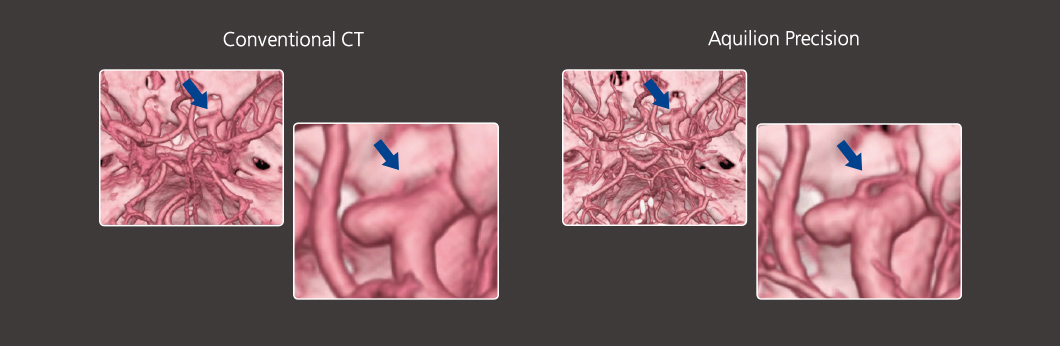
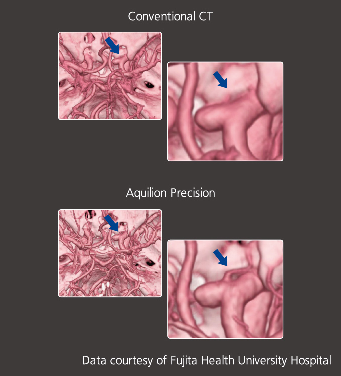
Comparison of CT images of cerebral aneurysm and ophthalmic artery (from same patient)
Since 1986, CT systems have been capable of resolving images up to 0.35 mm. Progress in visualizing finer detail was hindered by the fact that resolution could not be improved without corresponding increases in radiation dose to the patient. However, Canon Medical made a breakthrough in 2011 with the development of AIDR 3D, a technology that made it possible to take images with reduced radiation dose. Canon Medical then set about implementing this new technology in all of its CT systems. It also significantly improved two principal units—the X-ray tube and X-ray detector—to enhance resolution without increasing the required radiation dose. The result was the Aquilion Precision, which with a resolution of 0.15 mm more than doubled the resolution possible previously.
Canon Medical has also developed new technology that enables radiographers to adjust the size of the X-ray tube’s focal spot. By subjecting the electron beam emitted to the target for X-ray generation to an electric field, the electron beam can be narrowed by more than half, resulting in greater focus control. Applying an electric field in this way is like creating an “electrical lens” in the X-ray tube. Moreover, to improve the resolution of the X-ray detector, newly developed ceramic materials and machining processes were utilized, enabling a reduction in pixel size to one quarter that of the pixels found conventional X-ray detectors.
Looking ahead, Canon’s optical, image processing, and manufacturing technologies will continue to find synergistic applications in improved detectors, image processing and manufacturing technologies, as well as in advances in software for assisted analysis of high-resolution image data from CT scans and AI-based medical diagnostic support. These advances, made possible by combining the strengths of Canon and Canon Medical, will open up a wealth of new possibilities.
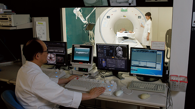
A clinical evaluation of the Aquilion One CT system.
Photographed at Fujita Health University Hospital