Technology supporting early detection of bone metastasis
If detected late, bone metastasis can significantly impacts patients’ quality of life. Canon is making it possible to visualize bone metastasis, which is difficult to detect early.
July 30, 2020Featured Technology
Challenges faced on the front lines of medicine
Cancer is a disease capable of spreading to different parts of the body. Bone metastasis--the invasion of a cancer into bone from a different, primary source--is difficult to spot unless the patient feels pain, making early detection difficult. Late detection or treatment of metastasis to the spine and other body parts that are of particular importance may also result in impairment of motor function, thus compromising the quality of life of the patient.
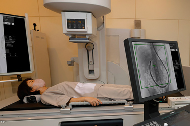
Typically, special tests such as bone scintigraphy are used to identify bone metastasis. This test utilizes a special camera to detect and capture images of radioactive substances administered intravenously to the patient. However, the procedure places a heavy burden on both the patient and the doctor as it is time-consuming and complex.
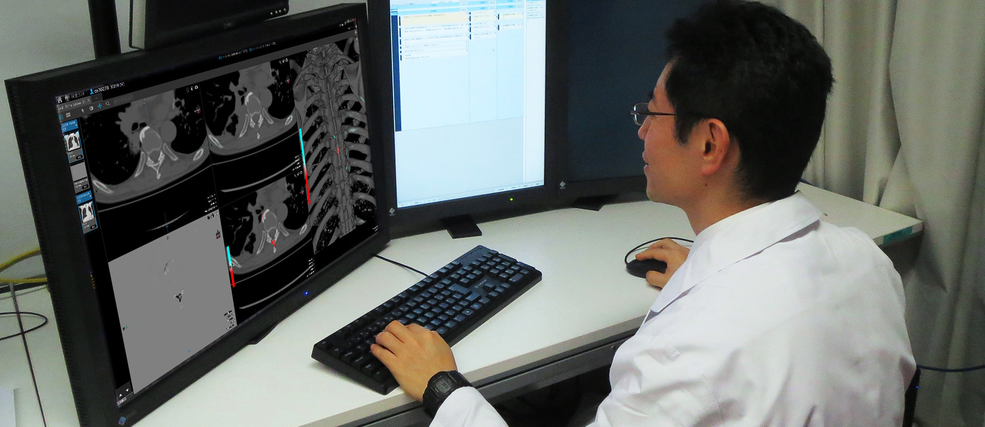
Canon has been engaged in various joint research programs together with Kyoto University Hospital. One such program is the development of a diagnosis support software program to detect the bone metastasis at an early stage. The decision to develop this software stemmed from the concerns of the doctors that came to light during the course of the frequent communication with developers at Canon. The standard practice in cancer treatment has been to determine the progression of cancer by studying images captured using computed tomography (CT). Identifying bone metastasis requires that past and present images be compared side-by-side to find changes that suggest bone is shifting. As CT technology has improved, the number of image slices has increased and bone can be observed in most of the image slices. However, time constraints typically afford only enough time to examine internal organs, and not enough time to fully inspect bone.
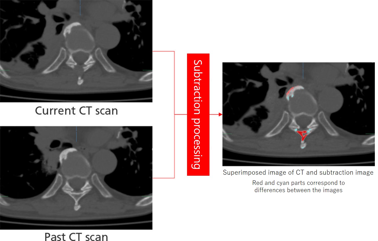
That’s where Canon decided to focus on. The company’s technology uses computers to compare previous and current CT images and subtract the difference. This not only helps prevent changes from going unnoticed, but also speeds up the inspection and reduces the workload of medical experts. Most importantly, doing so enables early detection and treatment of bone metastasis, thus contributing to improvement in the quality of the patient’s life.
A technique requiring more than simply aligning images
Detecting differences between the past and present images is not simply a matter of subtracting two images. Even with images taken of the same parts of the same person, a person's bodily conditions can change and cause tissue to be imaged differently. In the case of a cancer patient, for example, there may be changes in the body shape such as slimming and breathing difficulty. Thus, technologies are needed that can align images with great precision.
Besides bone metastasis, many other changes also show up in the subtracted image if we simply align the position of the two images. For instance, the difference may vary significantly even with how we breathe in while scanning. Canon therefore devised its own solution capable of producing subtracted images of the changes to bone even if the images are captured under different conditions.
First, a process is run to identify the bone region prior to aligning the images. Doing so makes it easier to grasp changes in the bone. Result will not be stable if fine-tuning the alignment is performed at the beginning, as this instability will slow down the process.
For this reason, a rough alignment is first performed by regarding the body as a rigid body (a body that is negligibly or not at all deformed). Image processing is then used to align the images—first performing a rough alignment, then gradually making more detailed adjustments.
Finally, a process is then performed to eliminate the remaining signal noise from the image thereby succeeding in generating a subtracted image that clearly depicts only changes in the bone, even for images captured under different conditions.
Bringing ONE CANON to the market
Canon, together with doctors, repeatedly carried out tests of data from more than 1,000 cases in the records of Kyoto University Hospital. The company eventually succeeded in developing technology that supports for the detection of bone metastasis. This was then included as application software for the “Vitrea” medical imaging workstation sold by Canon Medical Systems, which became a member of the Canon group in 2016.
Vitrea software is a multi-modality advanced visualization system providing comprehensive applications in a variety of IT environments. At present, there are more than 5,000 Vitrea systems currently operating in medical facilities around the world. The system’s software delivers high quality—including processing speed, precision and operability demanded by professionals. During the commercialization process, Canon and Canon Medical formed a joint team to further improve the usability of application software for searching possible bone metastasis from the perspective of end users.
Advance solutions were implemented in order to superimpose images of parts of the patient’s body to make bone metastasis more easily identifiable. While such procedures as image processing for torso CT images took several hours to complete, results can now be achieved more than 10 times as quickly.
Open innovation for further advancement
In the medical arena, challenges faced on the frontline cannot be resolved simply with the use of technology. Similarly, this technology that supports search for bone metastasis from cancer would not have been developed without open innovation in joint effort with Kyoto University Hospital. We are currently engaging in research and development work together with the said institution to further upgrade the current version.
If Canon is successful in applying this technology to other organs, the burden on both patients and doctors can be significantly reduced.
The entire Canon group will continue striving to make contributions toward further progress in medical care.