
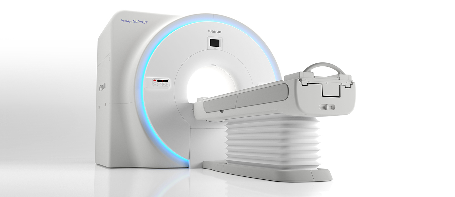
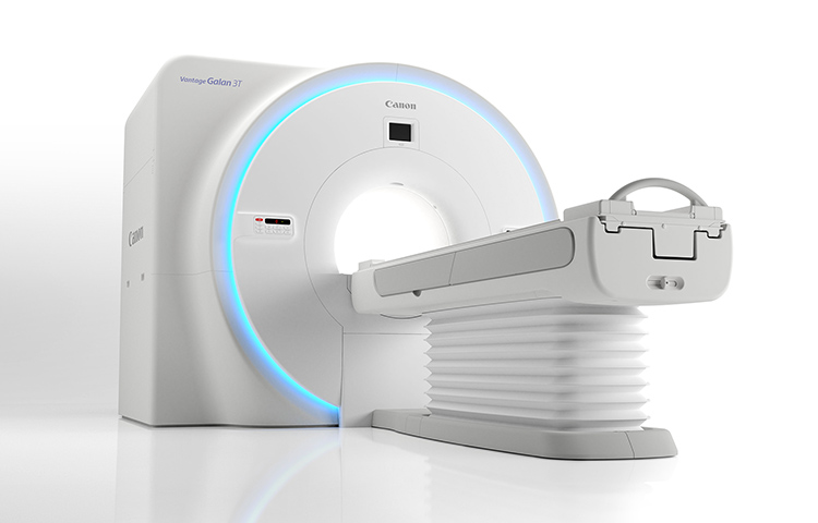
Magnetic Resonance Imaging (MRI) equipment makes it possible to obtain images of freely selected vertical, horizontal and diagonal cross sections of the human body using magnetic fields and radio waves. Because there is no risk of X-ray exposure and MRI scanner imposes little burden on the body, it can even be used to examine children without worrying about harmful effects. In addition, MRI scanner makes it possible to obtain clearer images of the brain, spinal cord and similar tissues compared with equipment that uses X-rays.
2018/12/27Featured Technology
#Pioneering Technology#Medical
Approximately 70% of the human body consists of water, which includes protons (hydrogen atoms). These protons are usually oriented in various directions. However, when they enter a strong magnetic field (a static magnetic field), they become polarized and face the same direction. If radio waves are then applied, the orientation of all the protons can be changed at once (magnetic resonance), and the protons revert to their usual state after some time passes.
MRI scanner utilizes this principle to make it possible for data collection/processing equipment and control equipment to output images of differences in the amount of energy generated by protons when they revert to their usual state. It is then possible to distinguish between the brightness of images of areas where the protons do not revert to normal very quickly (lesions) versus normal areas. MRI is therefore especially suitable for examining such soft tissues as those in the brain, spinal cord and torso. Images of blood vessels can also be easily obtained.
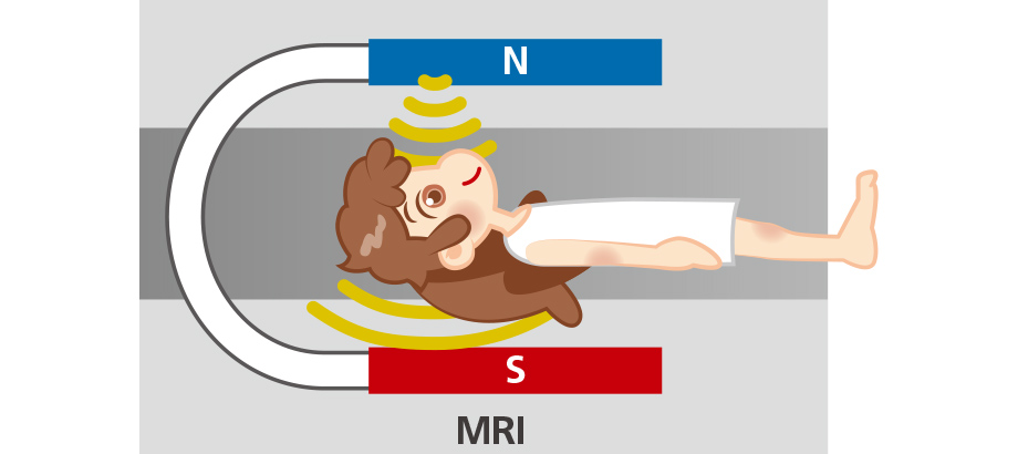
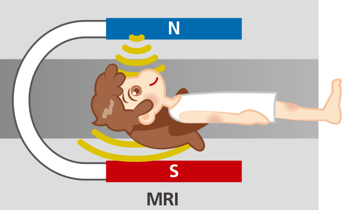
An illustration of MRI scanner. Real MRI scanner does not actually use a horseshoe magnet. Instead, MRI scanner uses a superconductive magnet similar to that used by a linear-motor-driven vehicle.

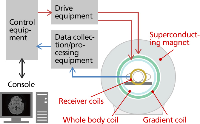
Structure of MRI Scanner
Although MRI is said to impose little burden on the body, there are also issues associated with MRI, including long examination times, the need to administer a contrast agent and the generation of acoustic noise. In order to reduce the burden on patients and shorten examination times, Canon Medical Systems is pursuing technological innovations to make possible examinations that are faster, more reliable and more comfortable.
#Healthcare#Social contribution#Patents#Electrical engineering
During an MRI scan, a lot of acoustic noise is generated, which can make patients uncomfortable or scared. This noise is caused by the current flowing through the gradient coil inside the large MRI magnet. When this current flows, force is generated in accordance with Fleming's left-hand rule, which causes the gradient coil to vibrate. These vibrations then reverberate to such parts as the magnet itself and cause the equipment to generate acoustic noise. However, with the PianissimoTM structure, Canon Medical's acoustic noise reduction technology, the gradient coil are vacuum-sealed to reduce the noise propagation by 90%. This makes examinations quieter and reduces the burden imposed on patients.
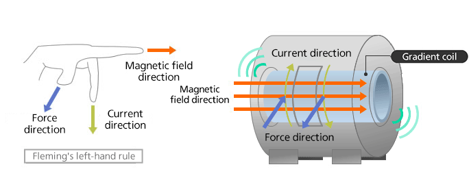
How the Vibrations that Cause Acoustic Noise Are Generated
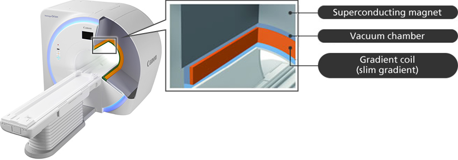
Development of a Vacuum Chamber to Seal Off the Gradient coil
#Healthcare#Imaging technologies#Social contribution#Mechanical engineering#Electrical engineering#Computer science#Physics#Chemistry
MRA (MR Angiography) refers to the scanning of blood vessels using MRI scanner. When a contrast agent is used to observe blood vessels, the obtained images are clear, but contrast agents can also cause side effects that include nausea, feelings of unwellness and headaches. Canon Medical started developing non-contrast MRA applications in the 1990s, ahead of other companies around the world. Starting in 2006, the company incorporated its non-contrast examination system into all of its MRI scanner. This system uses Canon Medical's Time-SLIP (Time-Spatial Labeling Inversion Pulse) method, which makes it possible to magnetically tag blood that you want to observe and scan only the target area.
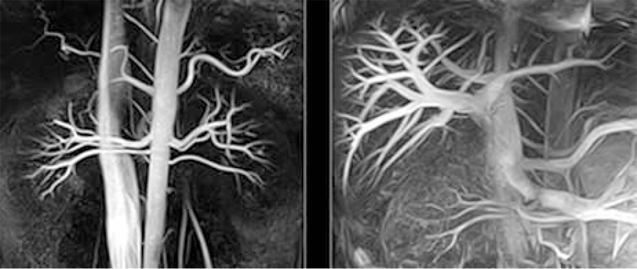

The aorta and renal artery (left) and vena cava and portal vein (right) as selectively depicted using the Time-SLIP method
#Healthcare#Imaging technologies#Social contribution#Automation#Mechanical engineering#Electrical engineering#Computer science#Physics
Because the heart constantly moves, it is necessary to accurately determine the position of a cross sectional view of the heart to examine it. To achieve this, patients have to repeatedly hold their breath, which imposes a burden on them and takes a lot of time as well as demanding the involvement of a skilled technician. To more smoothly start a heart MRI examination, it is important to shorten how long it takes to determine the position of the cross sectional view and simplify the process. Canon Medical has expanded its functions for assisting with determining the positions of cross sectional views necessary for diagnosis in order to achieve both examination efficiency and reproducibility.
This technology infers the position of the heart by comparing a scanned image with a model. The system's anatomical-structure recognition technology provides assistance with the bed position and volume-of-interest (VOI) settings during heart MRI examinations, which require expert knowledge. The equipment automatically detects the position of the subject's heart.
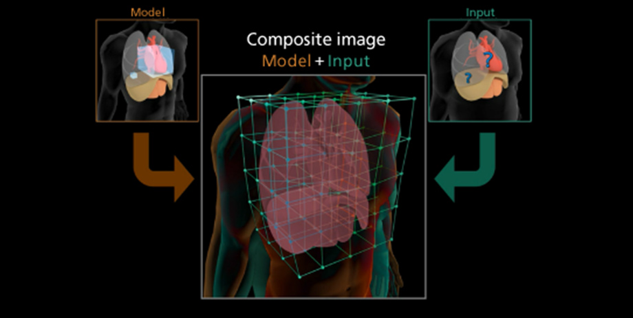
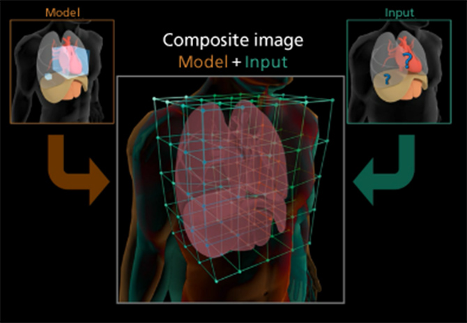
The scanned image is compared with a model, and the position of the heart is detected through the recognition of three-dimensional structures.
CardioLine+ uses example-based part inference technology that recognizes the statistical patterns of multiple distinctive parts of the heart to automatically infer the position and orientation of the heart regardless of the subject's particular physique, gender or medical conditions. This makes it possible for the system to accurately determine the positions of standard cross sectional views for heart MRI examinations. As a result, it is no longer necessary to repeatedly scan the patient as they hold their breath to determine the position of these views. Instead, 14 cross sectional views of the heart can be detected with just one scan of the patient while they hold their breath. These views include basic left ventricular views (the horizontal long-axis view, vertical long-axis view, left ventricular short-axis view, left ventricular four-chamber view, left ventricular two-chamber view and left ventricular three-chamber view), basic right ventricular views (the right ventricular short-axis view, right ventricular four-chamber view, right ventricular two-chamber view and right ventricular three-chamber view), the aortic valve, the pulmonary valve, the left ventricular outflow tract and the right ventricular outflow tract.
During a typical heart MRI examination, approximately 43%* of the examination time is used to perform such manual operations as setup. However, Canon Medical's assistance technology—which includes SURE VOI, a system that uses anatomical-structure recognition technology to automatically detect the position of the heart—and CardioLine+, reduce manual operation time during examinations to 9%, dramatically shortening the examination time. This makes it possible to greatly reduce the burden imposed on patients.