Technology in ProductsOptical Coherence Tomography
Early Detection of Eye Diseases
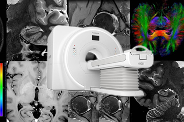
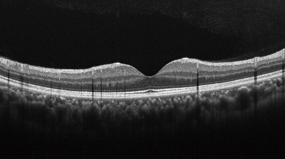
Optical coherence tomography (OCT) takes tomographic images of the retina and contributes to early detection and in follow-up checks of various eye diseases, including eye abnormalities due to diabetes and glaucoma. Light is controlled at the level of micrometers through the use of lenses and mirrors. The “wave” nature of light is used to take high-definition tomographic images.
March 23, 2023

How Optical Coherence Tomography Works
What is OCT?
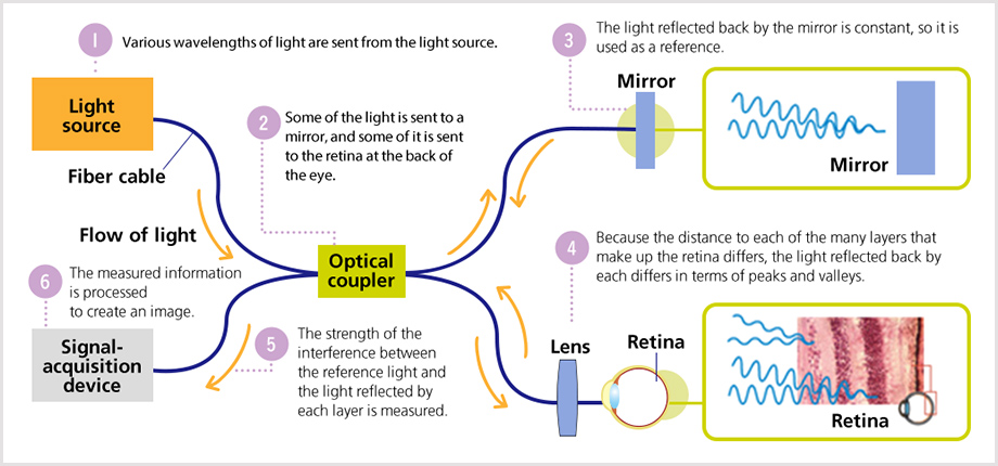
Light has “wave” properties, and "interference" refers to the phenomenon of the strengthening or weakening of waves of the same frequency when they overlap. Optical coherence tomography uses light interference to take cross-sectional images of the eye. It can observe, photograph, and record the ocular fundus and tomographic images of the fundus, and it is used for early detection and follow-up of ophthalmologic diseases such as age-related macular degeneration and glaucoma, which cause abnormalities in the retina and surrounding areas.
Read More
Canon Technologies Make This Optical Coherence Tomography Possible
Canon is helping to improve the accuracy with which ophthalmologic diseases are diagnosed by offering optical coherence tomography systems that are easy to operate and that quickly provide low-noise, high-quality images.
Optical Coherence Tomography Angiography (OCTA)
Canon’s optical coherence tomographies are equipped with an OCT Angiography (OCTA) feature that obtains images of blood vessels from OCT images.
OCTA technology depicts changes in OCT signal intensity over time to produce a high-definition image of blood flow. Contrast agent can cause a strong allergic reaction in some patients. However, OCTA does not use contrast agent, so it can determine the condition of blood vessels without burdening the patient’s body.
OCTA takes multiple OCT images at intervals at the same location. When these tomographic images are compared, the signal intensity changes because red blood cells are moving only in areas where there are blood vessels. By identifying the areas where the signal intensity has changed and selecting and highlighting those areas as blood vessels, an image can be obtained of fundus blood vessels similar to a fundus photograph taken using contrast agent.

An OCTA image of the fundus
OCTA Averaging
“OCTA averaging" is a method of reducing noise in an image by superimposing and averaging OCTA images.
Background noise is reduced by averaging multiple images taken at the same location. Then, distortion is corrected to yield high-contrast OCTA images.

Intelligent denoise (AI Noise Reduction)
“Intelligent denoise” is Canon’s unique new image processing technology using deep learning. Intelligent denoise removes signal noise from OCTA images, resulting in high-definition OCTA images that depict blood vessels in detail. Images from OCTA averaging are used as training data for deep learning. Intelligent denoise can produce high-definition OCTA images comparable to OCTA averaging, with just a single scan.


Wide-Angle OCT Imaging with Canon’s Unique Tracking Technology
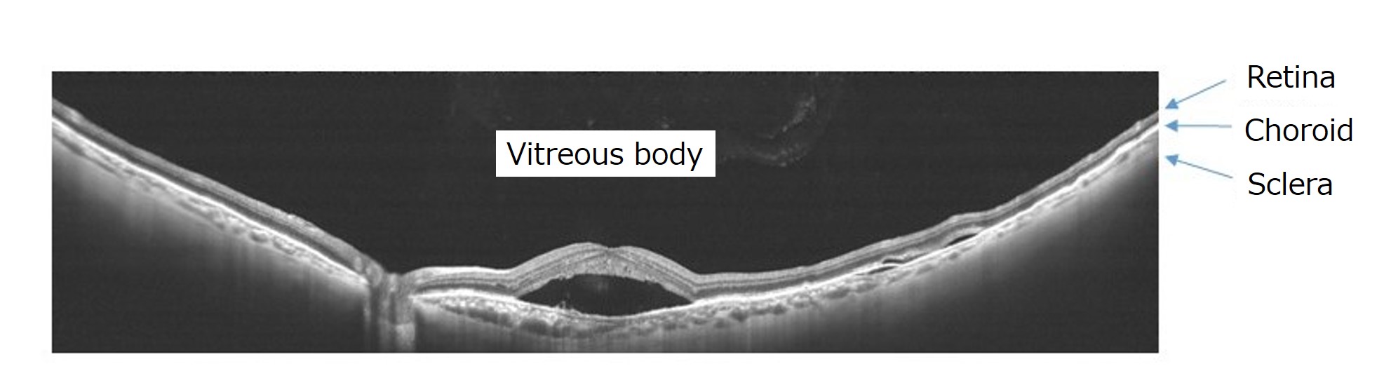
Swept source (SS) technology, which allows high-speed scanning, is used to provide wide-angle, deep imaging with a scan width of 23 mm and a depth of approximately 5.3 mm. An SS light source can quickly emit long wavelengths. Light can reach deep into the fundus, but the reflected light must be quickly processed into digital signals or else images cannot be obtained in a timely manner. A high-performance analog-to-digital conversion board is used to produce images via high-speed computing. In the past, tomographic images of a fundus with a long axial length (the length of the eyeball in the anterior-posterior direction), such as in cases of high myopia, could not be correctly rendered. Now, Canon’s technology allows high-resolution imaging of a vast area from the vitreous body to the retina, choroid, and scleral border in one session, enables the detection of retinal detachment and hemorrhage. Moreover, Canon has developed a unique tracking technology to reduce the effects of the subject’s eye movements and blinking, reducing the burden of imaging on the patient.
Canon’s Unique Image Processing Technology Enables High-Definition Wide-Angle OCTA Images
Wide-angle OCTA imaging of approximately 80 degrees* allows the identification of vascular abnormalities (such as nonperfused areas and neovascularization) due to conditions including diabetic retinopathy and arteriovenous occlusion in one session, greatly reducing the burden on the subject compared to conventional panoramic imaging. Moreover, “Intelligent denoise” is also used in wide-angle OCTA. This allows the acquisition of high-definition images. These images cover a wide area and allow the condition of capillaries to be checked.
*Horizontal 78°, vertical 68° (23 × 20 mm)

A high-definition wide-angle OCTA image that also allows the condition of capillaries to be checked