Technology

image

Minimizing the risks associated with radiation exposure and contrast medium to enhance patient safety and comfort

High-Definition CT System Revolutionizes the Field of Cardiology

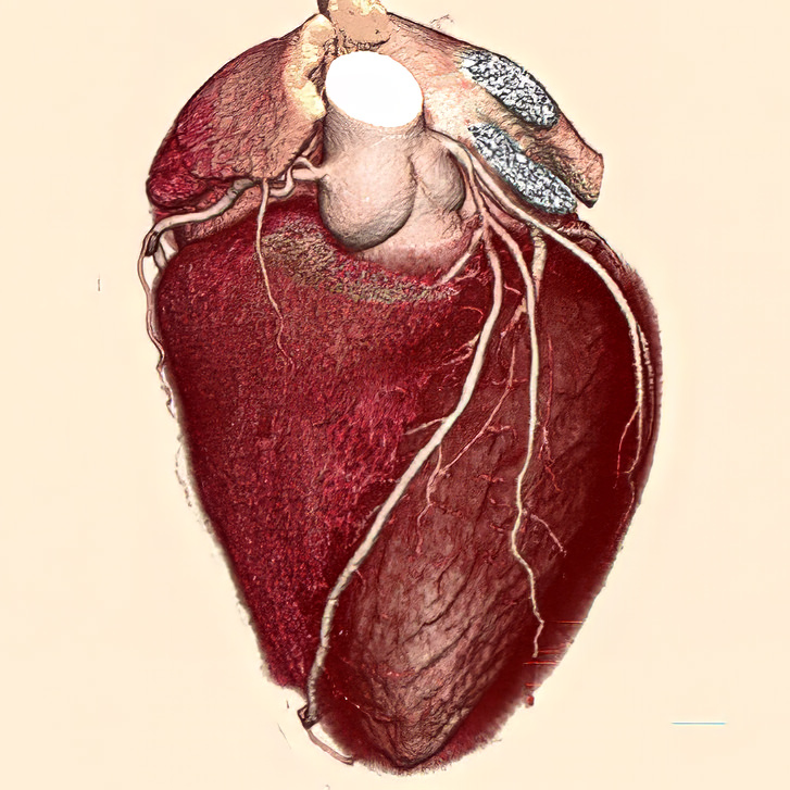
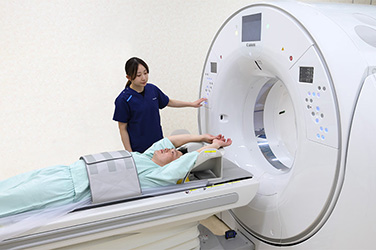
Canon's high-definition computed tomography (CT) system opens up new possibilities in diagnostic imaging by providing high-resolution clinical images. Canon has achieved high-definition, high-speed imaging capabilities that exceed all expectations based on joint research into new CT technologies in partnership with Johns Hopkins Hospital in the United States. Canon is dedicated to improving the health of individual people and of society as a whole by ensuring more accurate diagnosis and reducing the burden on patients.

April 7, 2026

Efforts and aspirations of Johns Hopkins Hospital

Could you discuss why you selected Canon as a partner for conducting joint research into CT technologies?

Dr. Zadeh:
Since its founding in 1889, Johns Hopkins Hospital has focused on public health. So we're trying to advance the health of patients overall as a society. But to do so, we need partners in industry who can provide advanced technologies.

Dr. Lima:
As the starting point, we were looking for a technique for imaging the coronary arteries*1 without having to place a catheter in the patient's body. It was in 2002 when I saw a picture at the American Heart Association meetings, and it was a picture from Canon Medical Systems.*2 We immediately began talking about siting the first machine at Johns Hopkins in Cardiology. That's how the association began.

Dr. Zadeh:
In many cases, the goals of industry are different from the goals of academia. The main goals of industry are obviously economic. Our interest is public health. However, we've developed a cooperative relationship based on a mutual division of roles, with Canon providing innovative technologies and Johns Hopkins Hospital providing the research necessary to validate these new technologies, based on a shared philosophy of contributing to people's health. It's been a wonderful partnership over the last 20 years, where we've seen a mutual respect for each other’s goals.

  • *1: The major blood vessels supplying oxygen and nutrients to the cardiac muscle (myocardium).
  • *2: Known as Toshiba Medical Systems Corporation at that time, with the name changed to Canon Medical Systems Corporation in 2018.
Image
Joao Lima, MD (Cardiology)
Johns Hopkins Hospital
Image
Armin Arbab-Zadeh, MD, MPH (Cardiology)
Johns Hopkins Hospital

Challenges

As a cardiologist, what's the most important thing you look for in diagnostic imaging devices like CT systems?

Dr. Lima:
It's the ability to accurately visualize and quantify plaque,*3 which is an indicator of atherosclerosis that may cause a myocardial infarction. In the past, it was always challenging to deal with artifacts caused by cardiac motion, which can easily affect image quality by causing image distortion. We also need to consider the amount of radiation exposure during examinations. In catheterization, a thin tube is inserted into the patient's body and 100 to 120 mL of contrast agent is administered in order to obtain clear images. This can place a burden on the kidneys.

  • *3: A mass of cholesterol and other lipids on the inner wall of a blood vessel that can cause narrowing of the vessel lumen.

Strengths of the Canon CT System

What are the advantages of your Canon CT system?

Dr. Lima:
Canon's CT systems have two major strengths. The first is the wide coverage.
With a wide 320-row x 0.5 mm detector, the entire heart can be acquired in a single rotation, reducing the radiation dose to one-fifth that of conventional systems.
The second strength is resolution. A high‑definition CT system with a 0.25 mm detector—far higher resolution than conventional models—has also been developed, enabling accurate visualization of extremely fine coronary arteries. With this improved resolution, the amount of contrast medium required during catheterization can be reduced to less than half of what was previously needed, markedly reducing the burden on the patient.

Image
Clinical image of the heart reconstructed from data acquired in a single rotation by a 320-row CT system

Dr. Zadeh:
The Canon system allows us to acquire images quicker. Because high-definition images can be acquired in less than 1 second, artifacts caused by respiratory and cardiac motion are minimized. That's a great advantage for patients who have difficulty holding their breath due to respiratory disease or advanced age. In addition, the ability to complete examinations in a shorter scan time reduces the burden on patients and streamlines the workflow of hospital staff.

Benefits observed after clinical introduction

How has Canon's high-definition CT system proved to be useful in clinical practice?

Dr. Lima:
The ability to capture clear images of the coronary arteries without the use of catheters, which is physically invasive for patients, is a revolution in the field of cardiology. In the past, catheterization was commonly performed to make the diagnosis, but catheterization can now be limited to patients who are identified as candidates for balloon angioplasty*4 based on high-definition CT images. Minimizing the risks associated with radiation exposure and contrast medium administration is also a great benefit for women and young patients. Previously, many women were reluctant to undergo CT angiography due to concerns about the impact on fertility and pregnancy, and the fact that CT angiography in women has increased to the same level as in men is a significant achievement.

  • *4: A therapeutic procedure in which a catheter with a small balloon at its tip is inserted into a blood vessel and the balloon is inflated to expand the vessel and increase blood flow.

Future prospects

What are your future expectations for Canon, especially with regard to collaborative research and clinical applications?

Dr. Zadeh:
We're excited about photon-counting CT (PCCT) which will dramatically change the field of cardiac CT. We hope that Canon will provide us with the most advanced technologies in this field, which will lead to the realization of an ideal system that combines higher resolution and an expanded scan width while reducing the radiation dose.

Dr. Lima:
PCCT not only provides ultra-high-resolution images, but also allows the acquisition of myocardial tissue information. It will allow us to obtain advanced information that can't be acquired by conventional CT systems, such as blood flow volume to the myocardium and the presence of cardiac scarring*5 or infiltration.*6 We look forward to how PCCT can provide more information.

  • *5: A process of myocardial damage in which the damaged cells are replaced by fibrotic tissues, resulting in reduced cardiac function.
  • *6: A process in which abnormal proteins or malignant cells invade the myocardial tissue, which can lead to heart failure in severe cases.
Image

Recommended Articles

Read articles in ”Realizing Professionals' Ideals”