Technology

image

MRI systems continue to evolve, with AI technology helping to realize more patient-friendly healthcare

Aiming to Make MRI Accessible to Everyone

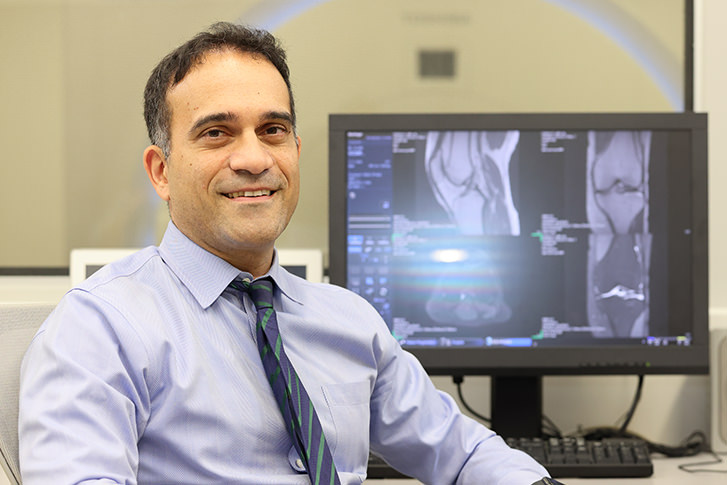
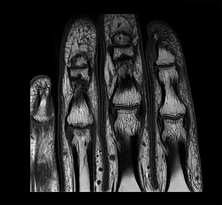
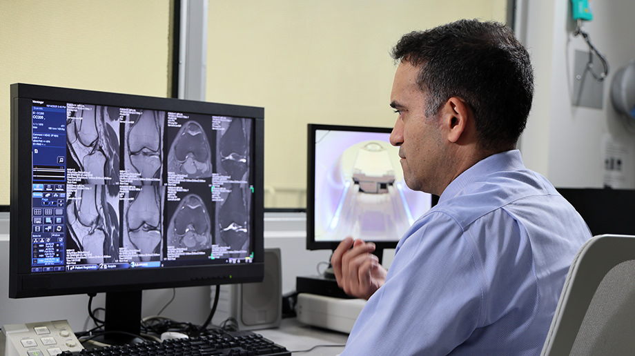
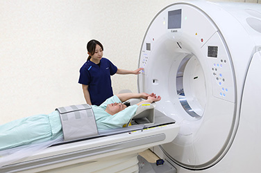
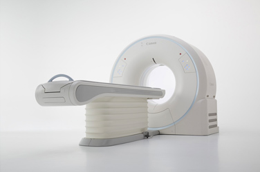
In the past, it was considered more difficult to achieve high image quality with magnetic resonance imaging (MRI) systems than with computed tomography (CT) systems. But thanks to recent technological advances, MRI is attracting greater attention as a diagnostic imaging modality that can play a major role in advanced medical care. Through joint research on MRI systems with Johns Hopkins Hospital in the United States, Canon is working to develop products that ensure a safe and comfortable examination environment for patients and provide new clinical value for healthcare professionals.

April 7, 2026

Efforts and aspirations of Johns Hopkins Hospital

Could you share your feelings about your joint research on MRI systems with Canon?

Dr. Demehri:
Canon is pursuing unique directions in MRI that other vendors aren't focusing on, such as "super resolution technology" for generating images with extremely high spatial resolution.*1 We feel that the joint research we've been conducting through our partnership, which has been ongoing since 2017, is extremely valuable. In the future, MRI systems will be employed in musculoskeletal imaging to diagnose and evaluate injuries and diseases of the muscles, bones, and joints. And MRI will contribute to medical care as a valuable alternative to examination methods that place a greater burden on the patient.

Dr. Lima:
The heart moves, which makes imaging difficult and leads to time-consuming examinations. MRI is an effective, and even indispensable, imaging modality for examining children and younger women, in whom the effects of radiation exposure are a particular concern. By utilizing AI, we aim to create a society in which anyone can receive MRI examinations easily and quickly.

  • *1: High resolution allows the detailed evaluation of extremely fine structures.
Image
Shadpour Demehri, MD (Interventional Radiology)
Johns Hopkins Hospital
Image
Joao Lima, MD (Cardiology)
Johns Hopkins Hospital

Challenges

What are the main challenges facing MRI, and are there any technological advances that can help overcome them?

Dr. Demehri:
In the United States, MRI of the knee is one of the most common imaging procedures for examination of the joints. But optimizing workflow, reducing scan times, and improving image quality have long been challenges. Several years ago, Dr. Lima introduced me to Canon's deep learning AI algorithm, and I was surprised and very impressed by the high image quality that could be achieved while reducing scan times.

Dr. Lima:
In the past, large or obese patients were considered unsuitable for MRI examination because the signals that are needed to generate images are weakened due to the greater distance between the coil*2 and the target region. But the introduction of AI technology has made it possible to obtain high-quality images of patients with any body type.

  • *2: During an MRI examination, a receiver coil is placed on the patient to increase sensitivity and obtain clearer images of the target region. Click here to view an article on the basic principles and technology of MRI.

Strengths of Canon's MRI system

What are the advantages of your Canon MRI system?

Dr. Demehri:
A great advantage is its ability to acquire images in shorter times with higher spatial resolution than before. This allows us to obtain high-quality images in a scan time of just 3 minutes. Furthermore, when combined with the fat suppression sequence,
*3 the whole brain can be scanned in less than 10 minutes, which is about one-third the time required with a conventional system. This is a major benefit for always-busy medical practices, increasing operational efficiency and cost-effectiveness. In the field of musculoskeletal medicine, we need to decide whether or not surgery is necessary by carefully evaluating structures such as small bones on a millimeter scale. Canon's MRI systems have extremely high resolution to support such clinical decision-making.

  • *3: This technology selectively suppresses signals from fat to more clearly visualize pathology such as inflammatory changes and tumors. Fat tends to show high brightness in clinical MRI images, which can make it difficult to observe lesions.
Image
Clinical image of the fingers obtained with a Canon MRI system

Benefits observed after clinical introduction

What are the benefits of Canon's MRI system for medical professionals and patients?

Dr. Demehri:
The greatest advantage is the ability to capture small nerves and joints more accurately by leveraging its high resolution. MRI scans performed using the Precise IQ Engine (PIQE)*4 and noise reduction algorithms jointly developed by Johns Hopkins Hospital and Canon can detect extremely subtle changes in the cartilage of the knee joint that could previously only be observed with an extended scan time. We were able to identify extremely mild damage in the meniscus of a study subject who was entirely asymptomatic, so we're confident that our MRI system will be useful for the early diagnosis of joint pain as well as for evaluating the progression of damage in asymptomatic patients.

  • *4: PIQE is used to reconstruct MRI images based on deep learning technology.

Future prospects

What technological innovations and clinical applications do you hope to see from Canon in our future joint research?

Dr. Lima:
The goal is to make MRI as accessible as CT. Since MRI examinations don't involve radiation exposure, they're extremely effective for people with heart disease, children, and younger women in whom radiation exposure is a particular concern. My main goal is to make MRI scans accessible to everyone by improving image quality and shortening scan times.

Dr. Demehri:
We're particularly interested in the development of post-processing tools employing AI for tissue characterization both in CT and MRI. Currently, in our lab at Johns Hopkins, we're developing a variety of algorithms to quantify tissues such as muscle, bone, and other structures. It would be a real game-changer for both patients and physicians if this function could be incorporated into Canon systems, so after an MRI scan of the affected area, the data could be automatically extracted and used as the basis for determining the need for a CT scan. This would minimize the additional cost and time associated with re-examinations. As a next-generation partnership between academia and industry, we're eager to make this happen.

Image

Recommended Articles

Read articles in ”Realizing Professionals' Ideals”