Technology in ProductsCT Scanner
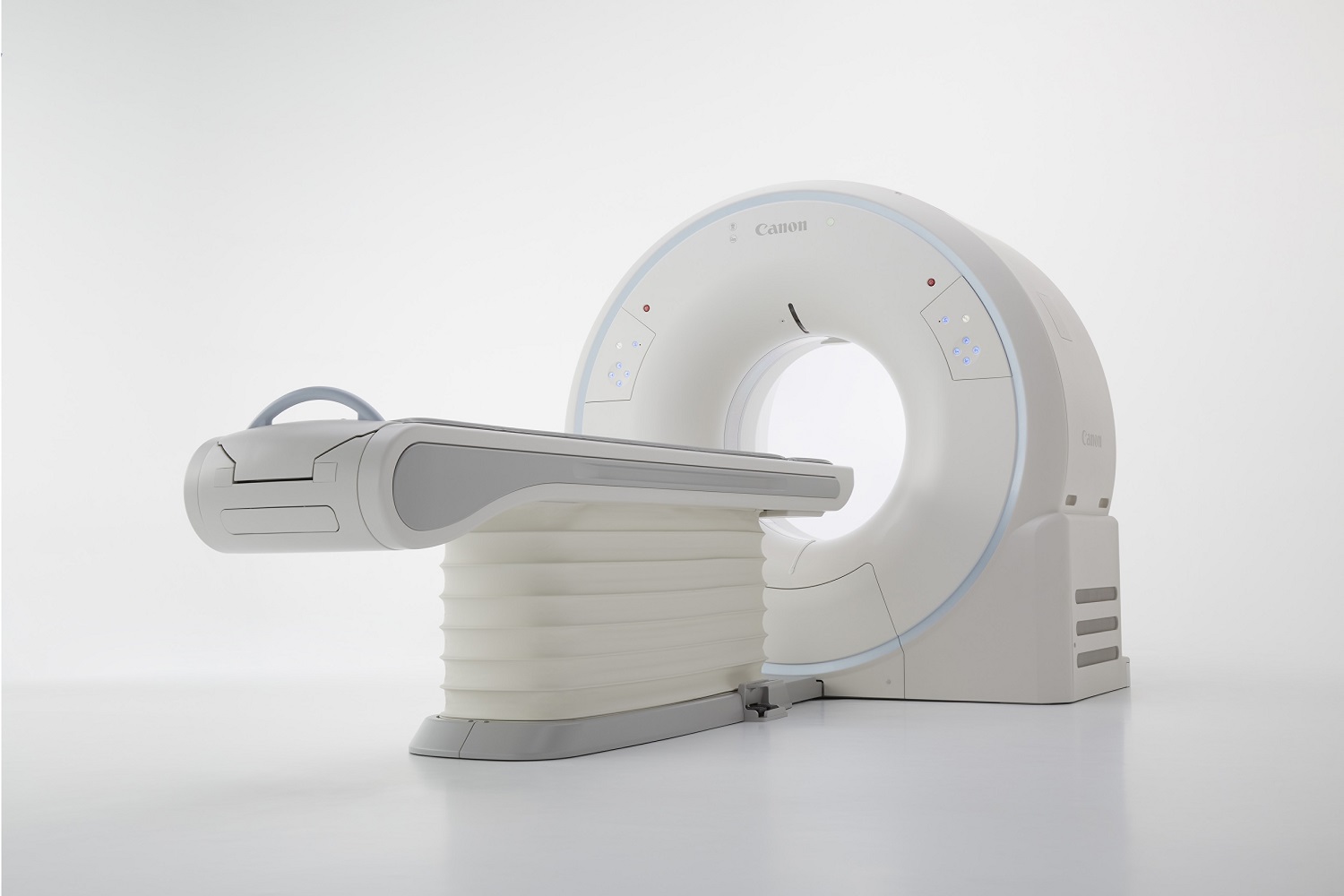
A diagnostic imaging device that uses X-rays to take cross-sectional images of the whole body
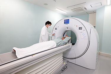
X-ray CT scanners use X-rays to take cross-sectional images of the whole body and are capable of detecting lesions that are just a few millimeters in size inside the body. Canon technology helps to detect disease early and to reduce the burden on patients while also reducing radiation exposure and imaging time.
March 21, 2024

How a CT Scanner Works
Ever-Evolving CT Machine
A computed tomography (CT) scanner is a diagnostic imaging device that uses X-rays. The gantry rotates an X-ray tube and an X-ray detector around a patient, and produces tomographic images based on the amount of X-rays through the body. Early X-ray CT systems, which appeared in the 1970s, used “non-helical CT” scanning. With each rotation, the scanner slightly shifted the X-ray tube and X-ray detector around a patient lying on the examination patient couch to take numerous images. Depending on the position of the cross-section, some "lesions” could not be found. In order to take higher quality images, “helical CT” scanning was developed. In this method, images are continuously taken in a spiral shape. Around 2000, "multi-slice CT” scanning was developed to obtain more precise images by increasing the number of images per rotation. With “multi-slice CT”, images are taken in a shorter amount of time with less radiation exposure. CT scanning has now advanced to "area detector CT" scanning, which can image the entire heart in 0.275 seconds.
Read More
Canon technologies in its CT scanners
In the development of CT scanners, it is essential to provide high resolution to detect small lesions in the body while also reducing the burden on patients. In order to achieve high image quality with a low radiation dose, Canon has developed technologies that image a wide area coverage and faster speed and more high resolution images.
320-row CT: Images of the heart and brain in one rotation
The greater the number images (slices) that can be acquired at one time, the shorter the examination time and the less burden is placed on the patient.
Canon has developed a CT scanner with a 320-row detector, allowing the acquisition of 320 images (slice width: 0.5 mm) per rotation. An area of 160 mm, along the axis of the patient’s body, can be imaged at one time. This enables the imaging of major organs of the body, such as the heart and brain, in a single rotation. But Canon’s CT scanner doesn’t just reduce imaging times.
Even if a patient’s organs change in size because of breathing or heartbeat, or if the patient moves during the imaging process, CT imaging can obtain a 3D image that seamlessly combines cross-sectional slices. However, the patient may still have to hold their breath several times depending on the area being scanned. This is a burden particularly for small children, the elderly, and severely ill patients. To make matters worse, CT scanners with fewer rows of detectors must acquire tomographic images of an organ in single slices, inevitably resulting in areas where the images overlap. Canon’s 320-row CT, however, can scan an extensive area in a single pass, significantly reducing overlapping. This results in an approximately 20% reduction of X-ray dosage for abdomen scans and approximately 80% for heart scans compared with conventional CT*, thus reducing the burden on the patient in terms of radiation dose.
*Compared to Canon’s conventional 64-row model
Low-dose imaging technology AIDR 3D
Generally, the X-ray dose required for a CT scan is proportional to the image quality.The higher the radiation dose the better the image quality.
In order to reduce the radiation dose given to patients while still maintaining image quality, Canon has developed Adaptive Iterative Dose Reduction 3D (AIDR 3D), a low-dose imaging technology that is standard on all of Canon CT scanners. Using various statistical models the noise in resulting images is reduced to approximately half of the conventional levels. High-quality images can thus be acquired without increasing the radiation dose given to the patients.

Example of a chest phantom with simulated nodules imaged with a low radiation dose (3 mAs)
Small simulated nodules which conventional reconstruction methods fail to depict are clearly visible
Advanced intelligent Clear-IQ Engine (AiCE)*, an image reconstruction technology based on deep learning
Canon has continued to develop a wide variety of technologies, such as AIDR 3D, which provides high-quality images while using low radiation doses.In 2018, Canon developed a new image reconstruction technology, called AiCE, which uses deep learning technology to further reduce the radiation dose and image noise while maintaining high image quality.
AiCE makes possible highly detailed scans with the same low dose radiation as that of a chest X-ray performed as part of a routine medical check-up.
AiCE is now being implemented in Canon’s CT scanners and is helping to reduce patient radiation doses while also helping improve physicians’ diagnostic abilities.
*A noise filter was used during the design phase of AiCE development. AiCE does not continue to learn once it is installed in a scanner.

A comparison of images obtained using conventional technology (left) and AiCE (right). Noise is reduced, resulting in higher image quality.


