High Resolution Images Acquired at Lower Exposure Dose Than Ever Before
Next-generation CT That Reduces the Burden on Patients
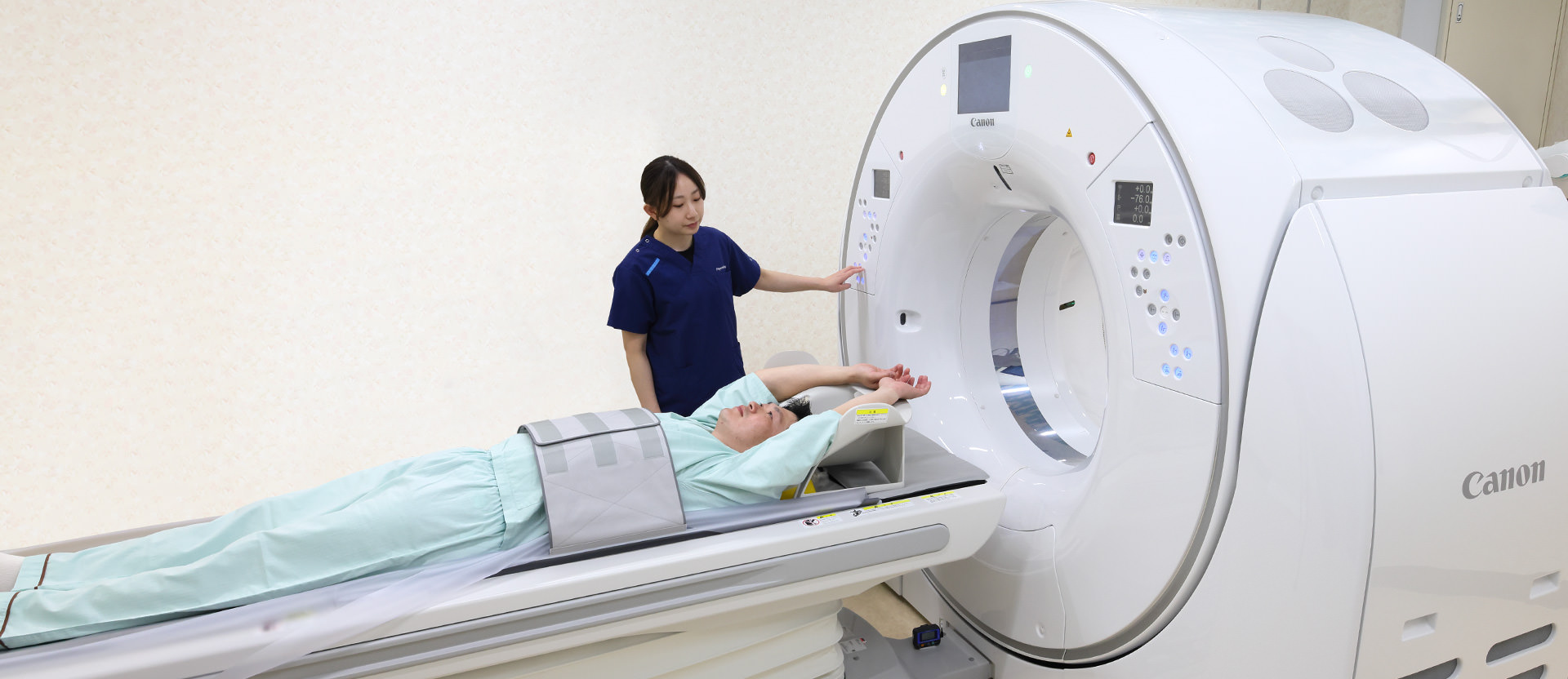
Hiroshima University Hospital and Canon Medical Systems (Canon Medical) are conducting joint research on photon counting CT (PCCT) to develop next-generation diagnostic imaging systems that can provide high resolution images at lower exposure dose than conventional CT. PCCT systems are also expected to be useful for more accurately evaluating tumor malignancy. A radiologist who has actually used a PCCT system comments, "I was amazed that the image quality was so good even when the exposure dose was reduced to only one-tenth." Clinical research is currently underway to fully realize this exciting new advance in medical care.
Hiroshima University Hospital is a leading medical center in Hiroshima Prefecture that makes great contributions to regional healthcare and provides the most advanced medical services. The hospital is designated by the Japanese Ministry of Health, Labour and Welfare as a special functioning hospital as well as a prefectural core hospital for cancer treatment and an advanced critical care and emergency center. One of the hospital’s core missions is "to pursue new medical innovations" by providing advanced medical care with the highest level of safety while focusing closely on their patients. In partnership with Japanese medical device manufacturers, the hospital is also conducting clinical research on advanced CT technologies to reduce exposure dose to patients and is actively involved in the dissemination of cutting-edge technologies from Japan to the rest of the world.
Diagnostic imaging systems are an indispensable part of advanced medical care. Dr. Kazuo Awai, Director of the Department of Diagnostic Radiology at Hiroshima University Hospital, is working together with Canon Medical to conduct joint clinical research on PCCT, which is a next-generation CT technology that can provide high resolution images at lower exposure dose than conventional CT.
Exploring the Possibility of Significantly Reducing X-ray Exposure
CT systems are used to examine many parts of the body, such as the brain, heart and blood vessels, bones, and so on. X-rays are generated by an X-ray tube that rotates around the patient, and the X-rays that have passed through the patient's body are received by a detector and sent to a computer in order to generate detailed tomographic images. As a general rule in CT examinations, the level of X-ray exposure is inversely related to the noise level in images. Increasing the X-ray exposure level can provide higher quality images, but at the cost of increasing the exposure dose to the patient. Conversely, reducing the X-ray exposure level can adversely affect image quality, resulting in images that are less clear.
PCCT, which holds great promise as the next generation of CT technology, is expected to overcome this tradeoff, making it possible to obtain high resolution images at an extremely low exposure dose. Dr. Awai notes, "Of the many advantages of PCCT, the most important is the reduction of exposure dose to the patient." Conventional CT systems generate diagnostic images by receiving many X-ray photons (the smallest quanta of light) and then performing measurement on the total number of photons that have been received. In this method, all the photons are measured together, so the measurement results include noise components. PCCT, on the other hand, is a faster and more accurate method in which each photon received by the detector is counted individually. Noise can therefore be identified and eliminated, resulting in higher quality images. PCCT makes it possible to obtain noise-free images at a much lower exposure dose. This is particularly useful for minimizing the burden on patients who need to undergo frequent repeat CT examinations to monitor the effectiveness of treatment or to check for disease recurrence.
Accurately Evaluating the Malignancy of Tumors etc. by Precisely Identifying Various Materials in the Body
In addition to its ability to precisely measure each individual X-ray photon, PCCT can also be used to perform multi-energy scanning by collecting and analyzing the energy information of each photon that has passed through various materials in the patient's body. This allows energy information related to specific materials in the body to be obtained with outstanding accuracy.
Dr. Awai says, "In conventional CT examinations, iodinated contrast agent is administered to the patient in order to identify the locations of tissues and lesions in the organs, but it is sometimes difficult to obtain good contrast with iodine when examining fine vessels or tissues and organs with low blood flow, such as those in the digestive tract. We expect that multi-energy scanning will allow us to quantitatively evaluate blood flow to various parts of the body and thus more clearly visualize conditions such as ischemia.* It may also be possible to identify whether or not tumors are malignant."
* A condition in which blood flow to an organ or tissue is reduced below the required level.
In PCCT examinations, it may be possible to administer multiple contrast agents simultaneously. The contrast agents used in conventional CT were limited to those containing iodine because it was not possible to discriminate between contrast agents with various compositions. However, because PCCT can identify substances based on their energy characteristics, it may be possible to simultaneously administer an iodinated contrast agent, which is difficult to deliver to areas with low blood flow, together with other contrast agents that can more easily reach such areas. Dr. Awai explains, "For example, in the diagnosis of thrombosis (a condition in which thrombus forms and leads to occlusion of a blood vessel), research is focusing on the visualization of atherosclerosis in all parts of the body by administering two types of contrast agents: one type that is taken up by thrombus and another type that remains in the blood. Substances that have never been employed before are now being considered as potential candidates for use as new contrast agents." PCCT is expected to be useful for evaluating lesions in areas that cannot be clearly visualized using conventional imaging methods.
Promoting the Dissemination of Advanced Technologies from Japan to the Rest of the World
Dr. Awai asserts that "PCCT has great potential in clinical practice." The performance capabilities of the PCCT system are currently being evaluated to obtain a clearer understanding of its practical value, and many new areas of research are being explored to validate its clinical potential. With regard to multi-energy scanning, we are also beginning to assess how the results obtained by this method can help to improve medical diagnosis. Dr. Awai strongly emphasizes the future potential of PCCT, stating, "There is no doubt that PCCT will improve the accuracy of diagnostic imaging, making it possible to see things that could not be visualized in the past."
Canon will continue to be actively involved in the research and development of PCCT, working together with Hiroshima University Hospital to create new clinical value and improve medical care.