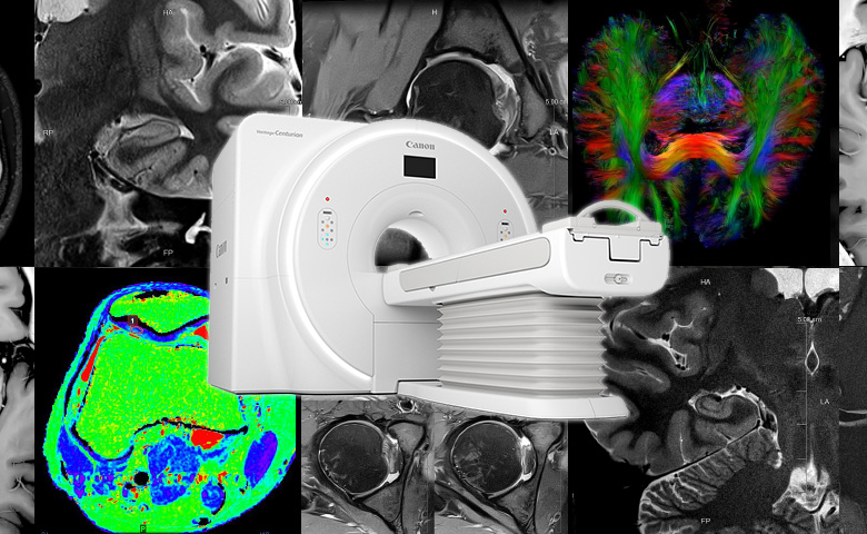
Deep Learning in MRI: Improved Image Quality, Shorter Examination Times
The Magnetic Resonance Imaging (MRI) is becoming widely employed as a diagnostic equipment that excels at the diagnosis of internal organs and muscles without using radiation. However, the long examination time has always been a heavy burden on both the patients and the medical institution.
At Canon, we have succeeded in employing the latest imaging technology that makes use of deep learning to enhance the image quality while shortening the examination time.
March 12, 2020Featured Technology
Removing the final hindrance to the growth of MRI
MRI and Computed Tomography (CT) scanners are two diagnostic imaging systems that many of us are familiar with, but do you know the differences between them?
The most significant difference lies in the signal source for obtaining the images. While CT scanners make use of high-frequency X-rays to obtain images, MRI do so using strong magnetic fields and electromagnetic waves with a low frequency that is equivalent to that of FM radio.
Both types of systems have their advantages, and are employed according to the intended purpose of the diagnosis or treatment. The MRI excels at the ability to detect lesions at parts of the body where X-rays are unable to produce a clear contrast, such as our brain, spinal cord, arm and leg muscles, and pelvic organs. As it does not subject patients to radioactive exposure, there is increasing demand for its use in a wide array of fields, including diagnosis, treatment and research.
However, MRI is also plagued by one issue in particular: Long examination times. An examination generally takes 20 to 30 minutes, but may sometimes extend beyond an hour. Not only does this inconvenience patients, the limited number of scans that can be done in a day prevents medical institutions from performing as many examinations as they would like.
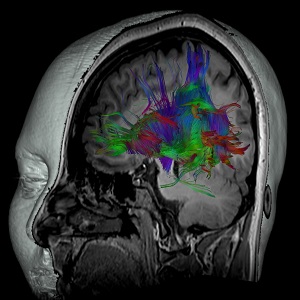
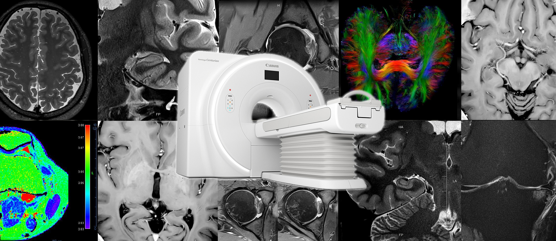
Cranial Nerves
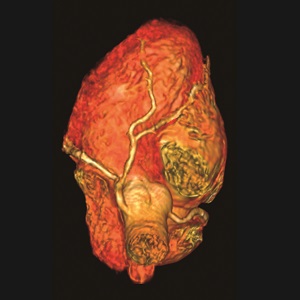
Cardiovascular System
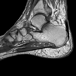
Ankle joint
The direction of the magnetic field and frequency of the electromagnetic waves can be adjusted on the MRI to capture images of different organs.
Trade-off between image quality and scan time
So why does an MRI take such a long time?
The water and fat content in our body contains hydrogen nuclei known as protons. An MRI forms images by collecting information from echo signals that are emitted when electromagnetic waves collide with the protons. Differences in the echo signals appear on the image in the form of contrasting tones. For example, the signal weakens and appears white at the places with no abnormality, while the signal intensity remains high and the image appear black at the places where a lesion is present. This contrast in the tone is an important feature of diagnostic imaging.
When the size of each pixel is reduced in order to enhance spatial resolution, it weakens the echo signal that is returned, which in turn lowers the contrast and makes it difficult to differentiate signals from noise. Overcoming this issue requires capturing images of the same spot repeatedly, which lengthens the examination time.
In other words, there is a trade off between higher image quality and a shorter scan time. This is what Canon’s group company, Canon Medical Systems (Canon Medical), is currently seeking to address.
Developing a system to produce images in 7T quality
To achieve both a shorter examination time and better MRI image quality, it is necessary to increase the magnetic field strength. Some MRI currently employed for general examinations offer a maximum field strength of 7 tesla* (7T). However, as they involve larger and heavier equipment and require a stronger, more robust scanning room, and enhanced magnetic shielding capabilities, they can be implemented at only a very limited number of medical facilities. As it is more practical for most of the facilities to install a 1.5T to 3T, Canon Medical has been devoting its efforts to develop a 3T that is capable of producing images in the 7T quality.
One approach that can make this happen is to enhance the strength the gradient magnetic field coil. In addition to the main static magnetic field, gradient magnetic field is also necessary for obtaining a cross-sectional image. Canon Medical has succeeded in strengthening the gradient field to enable capturing of images at a higher resolution than before. Not only so, it has developed an original high-speed imaging technique that involves capturing images at some instead of all data points, an approach that is also adopted in astronomical observation. At the same time, to minimize deterioration in the image quality caused by reducing the number of data points captured, Canon Medical has also created a unique algorithm to enhance both scanning speed and image quality. Yet another new development is the Advanced intelligent Clear-IQ Engine (AiCE), a reconstruction technology that makes use of deep learning to suppress noise.
- *Tesla: A unit of measurement for magnetic field strength
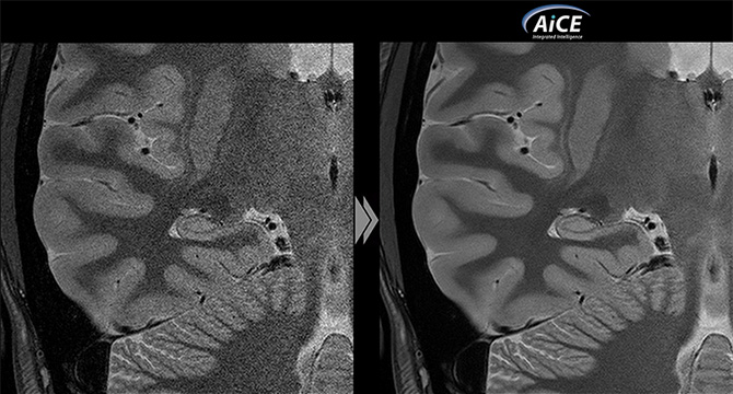
AiCE for MRI: The world’s first reconstruction technology to harness deep learning for noise suppression
AiCE is a deep-learning image reconstruction technology that is already available on Canon Medical CT scanners. Its implementation on an MRI is a world first. As CT scanners reconstruct images in a different way compared to MRI, AiCE for MRI was developed separately. Deep learning, an AI technology, was employed to reduce noise and obtain clear and high-resolution images in a short time.
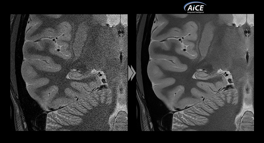
The development team was faced with many different challenges during the process. For example, diagnosis cannot be performed correctly if a part of the structure to be captured is erased together with the noise. There is also a need to train the neural network, which forms the basis of deep learning, so that it will retain structural details even when it removes noise. This training required a vast amount of image data with very little noise, and members of the development team went through countless photo shoots to capture about 30,000 images of their own bodies. These were then applied to the learning process to enhance the level of precision.
While the MRI is capable of producing clear images with minimal noise as long as imaging is performed over a sufficiently long duration, it is impossible for a single patient to go through such a long imaging process during the actual examination for various reasons. To make up for this limitation, Canon Medical developers spent extensive time capturing a massive amount of data for use as teacher images during the development of the AiCE technology for MRI. Also, a multi-layer neural network was constructed to process the signals used on MRI, which are different from those used by the AiCE technology for CT scanners.
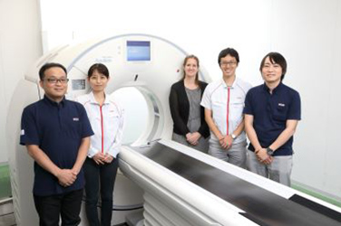
Highly rated by collaborating research institutes and hospitals
Before a medical device can be put into practical application, the new technology needs to undergo clinical trials to assess whether it is properly functioning and useful. Canon Medical has gained support from various collaborating research institutes and hospitals to verify the safety and usefulness under different scenarios. As a result, the technology has been rated highly not only for its safety, but also for aspects such as the shorter scanning time needed and the ease of diagnosis due to the reduced noise.
New prospects have also opened up with the high acclaim of the AiCE. For instance, in cases where an artifact appears in the image due to reasons such as movement of the patient’s body or magnetism, new possibilities have emerged, including application of AiCE in the technology to remove such artifacts.
Growing the market to contribute to the medical frontline worldwide
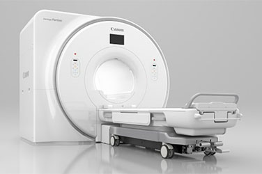
In Japan, Canon Medical released the “Vantage Centurian” in July 2019 and the “Vantage Galan 3T / Focus Edition” in October of the same year, both of which are MRI that are equipped with all of the above technologies. These systems aim at reducing the burden on the patient by shortening the examination time, enhancing the diagnostic performance with higher image quality, and improving work efficiency at medical institutions. Most importantly, it allows patients to receive more appropriate diagnosis and medical attention.
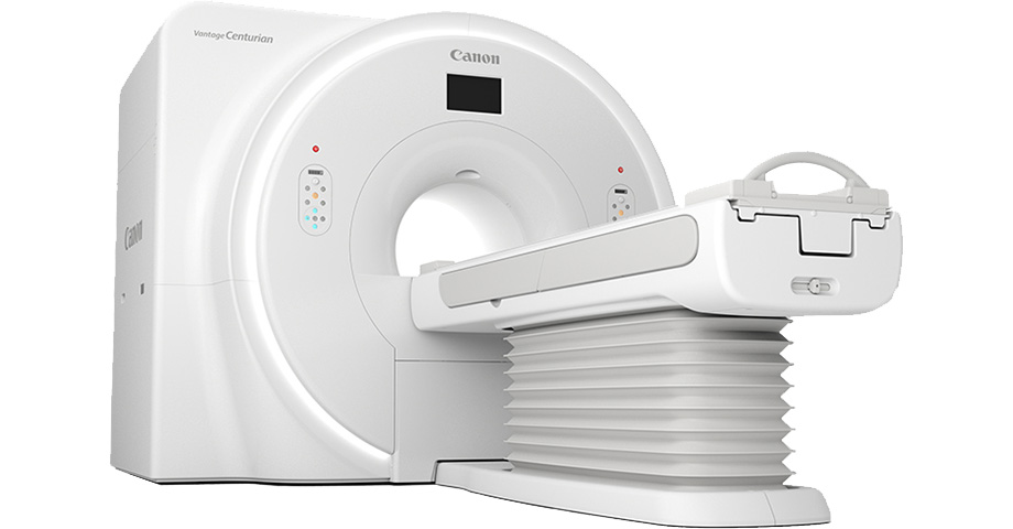
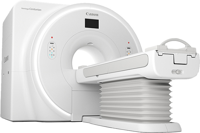
“Vantage Centurian” is the world’s first MRI to be equipped with a deep learning-based noise suppressing reconstruction system.