
Examination and treatment for a wide range of specialties
Pursue dedicated fluoroscopic image for diversified clinical needs.
Canon's digital fluoroscopy system brings high image quality, low dosage and unrivaled versatility to Tokyo Women's Medical University Yachiyo Medical Center (TYMC), a hospital with advanced medical services trusted and respected by the community.
October 13, 2022
Confident testing and treatment
In addition to being a university hospital, TYMC serves as a regional core hospital, general perinatal care center and pediatric critical care center. In 2021, following a rigorous review, TYMC replaced the multipurpose system in one of its two X-ray fluoroscopy rooms with a Canon Medical digital fluoroscopy system.

TYMC opened in 2006 and plays a vital role in the local community

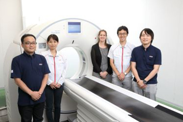
Mr. Michio Nakayama, Chief, Imaging Exam Room, Department of Medical Technology, TYMC
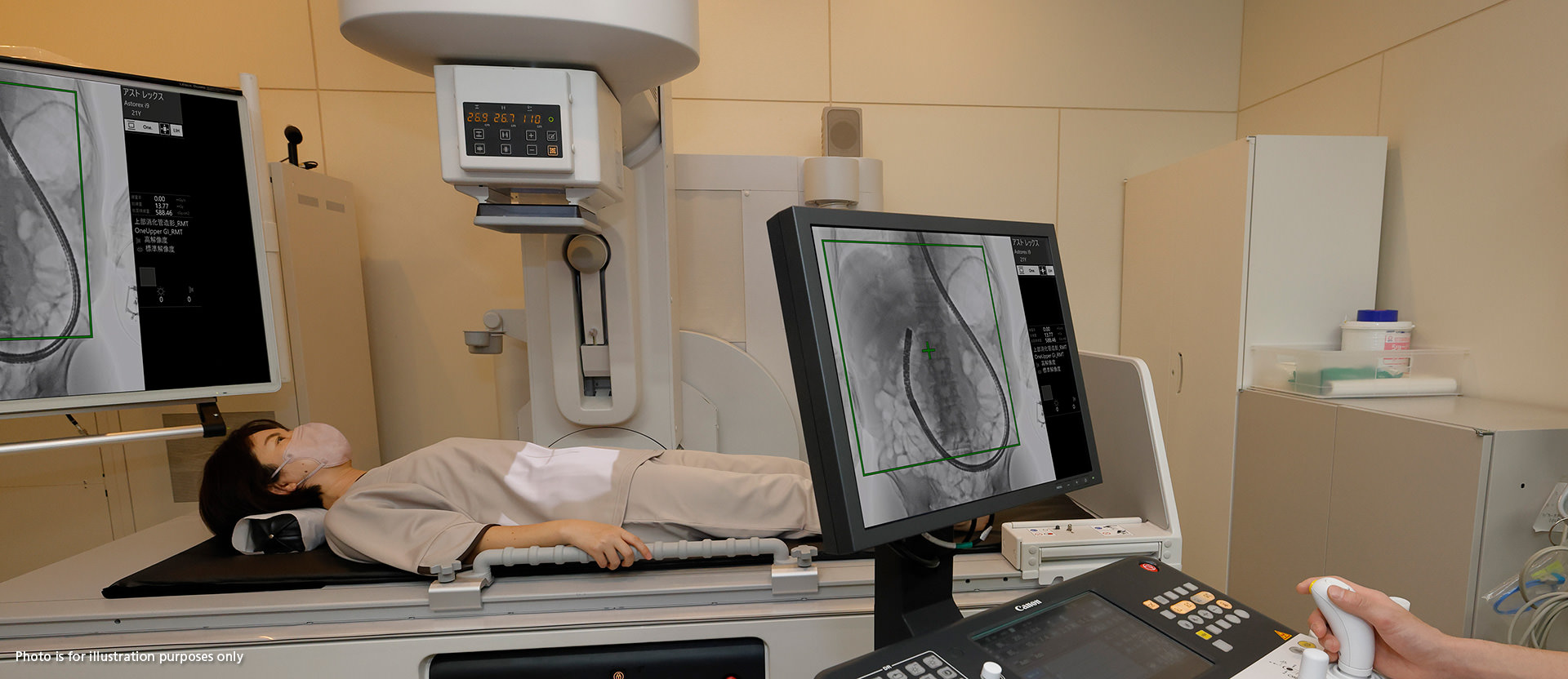
Digital fluoroscopy systems send X-rays through patients’ bodies to a flat panel detector (FPD) that captures still and video and images of internal parts for real-time fluoroscopic observation. These systems are mainly used for gastroenterology examinations and various urology, orthopedics and anesthesiology examinations and procedures. In recent years, advances in endoscopes and catheters have led to increased use of such devices for examinations and treatments in various clinical departments including fluoroscopy (where contrast agents are often used), gastroenterology (for digestive system surgeries, etc.), orthopedics, pediatrics and urology.
“We can confidently offer examinations and treatments in various clinical departments.”
According to Mr. Michio Nakayama, a radiologic technologist at TYMC, the hospital’s previous fluoroscopy system required movement of the patient when adjusting the field of view. This made it difficult to meet the varying needs of different clinical departments. However, Canon Medical’s digital fluoroscopy system simplifies examinations and treatments for all departments, because the field of view can be changed without moving the patient and high-quality images can be obtained with low radiation dosages.
High-quality images at low dosage
With the previous digital fluoroscopy system, image quality could only be improved by increasing the dosage and radiation exposure. If the dosage was reduced, details couldn’t be clearly identified.
“Doctors have praised the image quality of the new system. For example, when inserting a CV catheter* into a blood vessel in the chest, the catheter passes near the spine. Since the lungs are full of air, it’s usually difficult to match their image quality with that of bones because they require different dosages. But Canon Medical’s system uses image processing to adjust image quality and deliver high-contrast images in real time even when the dosage is suitably low for the lungs,”explained Mr. Nakayama.
- *Central venous catheter for nutritional supplementation when oral or enteral feeding is difficult.
By combining an all-new image processor with other proprietary technology including a 17x17 inch FPD, Canon Medical succeeded in reducing noise, improving contrast resolution and raising fluoroscopic image quality at dosages below what is required by conventional models.

The catheter can be clearly observed even in areas where viewing is usually difficult.

Myelogram using conventional fluoroscopy

Myelogram of the same patient using Canon Medical’s digital fluoroscopy system
Comparison of conventional fluoroscopy with Canon’s digital fluoroscopy system using images from the same patient
Canon Medical’s system uses about one-tenth the dosage to obtain image quality comparable to that of the conventional system.
Improving the test environment for safer treatment
The digital fluoroscopy system previously used by TYMC was bulky and wide, making it difficult for patients on beds and wheelchairs to enter and exit the exam room. The narrow working space was also a major issue because various equipment often had to be carried in with the patient.
“The compact wall-hugging design was a key reason for adopting the system. Once the system was installed, the examination room had much more available space, allowing us to flexibly place IV stands, endoscopes and ultrasound devices. In the past, there was so little space around the X-ray table that the doctor could only stand in a limited area. Now there is much more freedom of movement, allowing treatment even from the patient’s head or feet. The flow of staff members has also substantially improved, making it possible to perform treatment with greater peace of mind than before.”
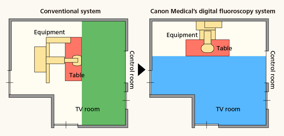
Canon Medical’s system improved the X-ray fluoroscopy room layout
The previous system was positioned parallel to the control room, which made it difficult to secure space in front of the X-ray table (green area) while also allowing medical staff to move. But Canon Medical’s system could be installed against the wall to create ample space in front of the X-ray table (blue area) for both the doctor and staff to move about.

The system is particularly compact in depth, allowing the doctor to stand to the left, right or front of the X-ray table in a natural posture at all times for safer treatment.
There is no need to move equipment to change the field of view
Conventional digital fluoroscopy systems can only magnify the central part of the FPD to enlarge the image while surgery is being performed. With Canon's new feature, it is possible to shift the field of view to any area within range of the wide 17x17 inch FPD to enlarge the image. Thus, there is no need to move the X-ray table or video system components such as the X-ray tube or detector. Additionally, the patient does not have to be repositioned.
“Even before we introduced the system, we thought this feature was in line with our desire to offer patients greater safety and peace of mind during examinations. But actual use showed the effect surpassed our expectations. Moving the table while a needle is in the patient’s body may cause the needle to come off. In such cases, we always asked the doctor performing the treatment to stop moving their hands before repositioning. Eliminating the need to move equipment also allows safer treatment, such as when replacing a tube in the flank during liver surgery. I feel it expands our options and encourages doctors to perform examinations."

Any area within range of the FPD can be enlarged and displayed without moving the X-ray tube or FPD. The area, in a choice of three sizes, can easily be selected using the joystick control lever.
Unlike conventional digital fluoroscopy systems that require mechanical movement of the X-ray table or video equipment, Canon Medical’s system is silent and vibration-free, which further benefits patients.
“Since patients’ field of view is often limited during examinations and treatments, the sounds and vibrations generated by moving equipment may make them feel nervous. Keeping the system stationary helps patients feel more secure.”
Moving forward, TYMC expects Canon Medical’s digital fluoroscopy system to play an even more active role in flexibly supporting various medical treatments. It also plans to use the dosage report feature to further optimize dosage management, reduce the dosage exposure of patients and more.
Generic Name |
Brand Name |
Certification No. |
Manufacturer and Seller |
Stationary digital general-purpose X-ray fluoroscopy diagnostic device |
Digital Fluoroscopy System Astorex i9 ASTX-I9000 |
302ADBZX00081000 |
Canon Medical Systems Corporation |


