
X-ray Angiography: Supporting Catheter Therapies through Real-time Fluoroscopic Imaging of Blood Vessels
Catheter therapies treat diseases in the blood vessels of the heart, brain and other parts of the body by inserting a fine tube called a “catheter” into a blood vessel. X-ray angiography, which enables real-time fluoroscopic imaging of the blood vessels, plays an essential role in catheter therapies. Canon offers an unprecedented level of high-definition image quality and operability in this area, which helps to significantly reduce the burden on patients.
December 23, 2020Featured Technology
Fluoroscopic imaging of the blood vessels in real time: X-ray angiography provides support for catheter therapies
An aging population and changing lifestyles have led to an increase in the frequency of such vascular diseases as heart attack and brain aneurysm. Amid this trend, catheter therapy is becoming increasingly preferred as it not only removes the need for major surgical procedures such as thoracotomies or craniotomies, which impose a heavy burden on the patient, but also helps to greatly shorten the length of patients’ hospital stay.
X-ray angiography is an essential part of catheter treatment. The procedure introduces contrast media into the blood vessels of the patient in order to obtain real-time intravascular images, which are displayed in monochromatic contrast. Originally developed for performing diagnostic examinations, it is now an important tool for medical treatment. In the emergency department—where every moment counts—X-ray angiography is even more useful as it makes the process from diagnosis to treatment more streamlined and speedy.
In addition to heart attacks and brain aneurysms, catheter therapy is also used to treat such conditions as varicose veins and arteriosclerosis in the foot, occluding blood vessels to prevent nutrients from reaching a tumor.
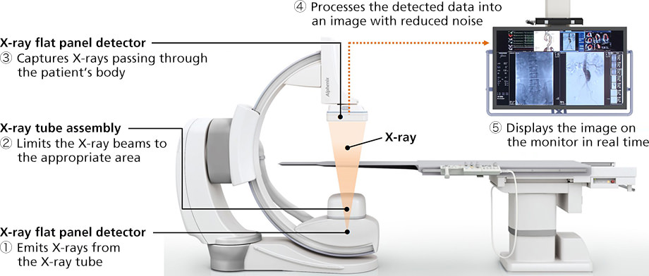
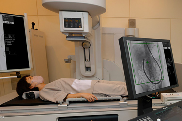
① Emits X-rays from the X-ray tube
② Limits the X-ray beams to the appropriate area
③ Captures X-rays passing through the patient’s body
④ Processes the detected data into an image with reduced noise
⑤ Displays the image on the monitor in real time
How X-ray angiography works
Pursuing high image quality to address diverse treatment needs
An X-ray angiography system acts like a surgeon’s eyes—it must display captured images on a monitor in real time and in high resolution. The latest system from Canon is equipped with an X-ray detector featuring a dynamic range (the range of brightness from the brightest to the darkest tones) 16 times wider than currently available systems. Switching from conventional 12-bit image processing to 16-bit processing enables the system to deliver images with higher concentration resolution. The result is a more refined gradation of the monochromatic contrast, making it possible to view fine blood vessels and tissues that could not be captured in the past.
Generally, an increase in the amount of information to process leads to a greater amount of noise in the resulting image. However, Canon has successfully overcome this trade-off by updating the flow of operations, from the generation of X-rays the displaying of images on the monitor, by incorporating a new proprietary noise reduction technique, improved processing and high-speed calculation that optimize the quality of images.
As different clinical departments have different requirements for image quality, Canon employs application specialists who possess relevant qualifications, such as “Clinical Examination Technician,” and gather feedback from frontline healthcare staff around the globe and report back to product development teams. Through this process, Canon is able to achieve the most appropriate level of image quality for a given treatment.
The world’s first high-definition detector for spotting fine blood vessels in the brain
While X-ray angiography can be employed for various treatments in areas of the body ranging from the brain to the feet, the necessary level of image quality varies depending on the method and region of treatment.
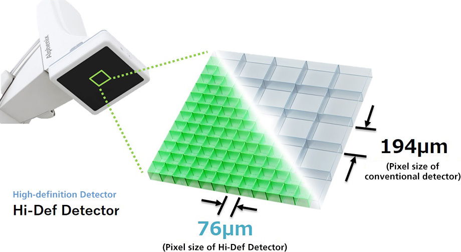
The brain, in particular, has an intricate network of fine blood vessels threading through the gaps between the nerves, and there has been significant demand from clinics at the point of care for an X-ray detector with a higher resolution. Through joint research with the doctors at the University at Buffalo, a distinguished institution in neurosurgery located in New York State, Canon succeeded in developing the “Hi-Def Detector,” a high-definition X-ray detector with a pixel size of 76μm—less than half the size of pixels on conventional standard detectors, and the world’s smallest pixel size among X-ray angiography detectors (as of September 2020).
One scenario in which high-definition detectors prove useful is the coil embolization of brain aneurysms. This treatment prevents rupture of the aneurysm by inserting coils into the blood vessel in order to block blood flow into the aneurysm. While the burden placed on the patient is less than that of other surgical treatments, there is a risk of relapse if the coils are not packed sufficiently. Canon’s high-definition X-ray detector is capable of producing clear images of the inserted coils without “crushed blacks” that erase details in darker areas. This technology has been highly praised by frontline medical staff, who commented that it allowed them to view the movement of the coils much more closely than before.
Comparison of a conventional detector (left, Digital zoom) with the Hi-Def Detector (right, High Definition mode)
*In order to view videos, it is necessary to consent to the use of cookies by our website. If the videos are not displayed, please click the "Cookie Settings" and accept cookies.
Shorter treatment time through safe, stable, low-vibration and high-speed operation
As X-ray angiography systems are employed onsite in operating rooms and emergency departments, in order to save lives, it is important to ensure that not only can the X-ray detector be used safely without coming into contact with other people or equipment, but also that it can be moved quickly and precisely into the desired position.
In terms of safety, X-ray angiography systems have a substantial weight imbalance due to the heavy X-ray tube and detector installed on the arm. As such, serious accidents may occur if the arm comes into contact with the patient. Contact with other examination or medical equipment nearby may also cause disruptions to the treatment. Canon’s system accounts for this issue by providing excellent operability with a well-balanced combination of different technologies, such as a safety mechanism to prevent object contact and a compact design for operation in environments with limited space.
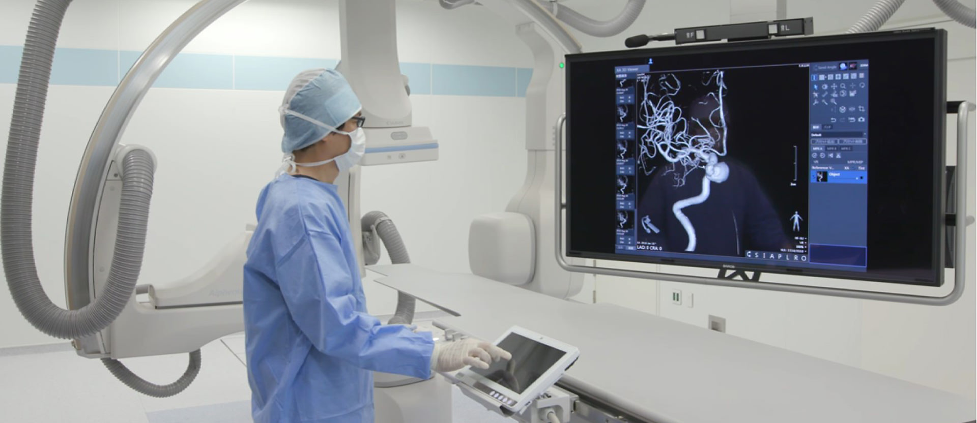
Meanwhile, in terms of speed, the ability to quickly move the arm to the intended working position and angle and immediately secure it in place is crucial to shortening treatment time. In 3D imaging, where images are captured while rotating the arm, imaging time is affected by the rotation speed. However, moving the arm at high speed increases vibration, resulting in a poor-quality 3D image. Highly sophisticated robotic technology is therefore required to make possible high-speed operation that minimizes vibrations. By working to suppress vibrations and developing original software for driving the system, Canon has delivered both high speed and stability.
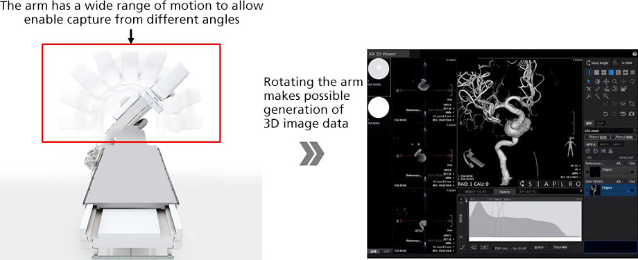
In 3D imaging, the system rotates the arm and utilizes the captured images to create 3D image data of the blood vessels
Visualizing radiation dose to safeguard patients and healthcare staff
Among the X-ray angiographic technologies that Canon has long pursued is the reduction of radiation dosage. Prolonged catheter treatment inevitably increases the level of radiation exposure that the patient is subject to.
The Dose Tracking System is a proprietary tool developed by Canon that reduces radiation exposure by generating visible X-ray beams, which are normally invisible to the human eye. By providing a color display of the incident skin dose and area of X-ray irradiation on a model image of the patient in real time, this technology enables catheter treatment to be performed while monitoring the level of radiation exposure. In addition, Canon has developed a proprietary technique that allows X-rays to be concentrated only on the necessary area, thereby helping to reduce the exposure of patients and medical staff to radiation.
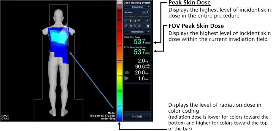
Dose Mapping Function
Dose Tracking System (DTS)
Catheter therapy helps to significantly alleviate the burden of medical treatment for diseases that conventionally required major surgery, and is likely to make further progress in the future. By addressing diverse needs, safeguarding health and protecting precious lives, Canon will continue to enhance its techniques for realizing its medical business philosophy of “Made for Life”.


