8K technology plays a part in Hokkaido University's cutting-edge 3D imaging system.
Revolutionizing Fossil Analysis with 100 Million Times More Information
Canon’s 8K imaging technology plays a key part in Hokkaido University’s 3D imaging system, which employs a new fossil analysis technique to help capture images of ancient life.
December 26, 2019Featured Technology
Contributing to next-generation 3D imaging technology that accelerates the research of evolution
Life on earth first began four billion years ago. Compared to such a great span, the history of mankind is but a fleeting moment. Many paleontologists have been trying to unravel the mystery of billions of years of evolution and they continue to make steady, gradual progress. However, there yet remain many unanswered questions about it.

Tomographic image of sample captured via CT scan
The study of fossils is crucial for understanding how life has evolved over such an enormous span of time. Numerous fossils can be found within rocks and strata on the surface of the Earth. In a way, they are a sort of “storage media” that contain a continuous record of history of life.
Dr. Yasuhiro Iba, an Associate Professor at Department of Earth and Planetary Sciences, Hokkaido University, is one of the researchers working to unravel the mysteries of evolution. His research team focuses on capturing tomographic images of rocks using high-performance digital cameras. The team has succeeded in developing an imaging technique that involves overlaying cross-sectional images, enabling more efficient, higher-resolution fossil surveys than through conventional approaches. An essential element of this technique is Canon’s 8K imaging technology.
State-of-the-art tomographic techniques using digital cameras
Many researchers make use of X-ray CT scanners when inspecting the interiors of fossils and rocks. While the use of X-rays allow researchers to survey samples without destroying them, it cannot produce detailed information about their internal structure. In addition, images are captured in grey scale with low resolution, and shown only in terms of light and dark contrasts, preventing researchers from obtaining accurate images of organisms that lived in the past.
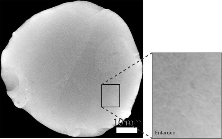
Tomographic image of sample captured via CT scan
Aiming to overturn conventional wisdom in the world of palaeontology that “non-destructive analysis” is the only viable technique, Dr. Iba believes it is possible to adopt a completely new fossil analysis technique using digital cameras. This technique involves polishing the surface of a fossil or rock sample, then capture an image of the sample using a Canon EOS 5Ds ultra-high-resolution digital camera (equivalent to approx. 9K resolution). After repeating the process more than 1,000 times, he successfully produced a sequential of tomographic images in full color and ultra-high resolution.
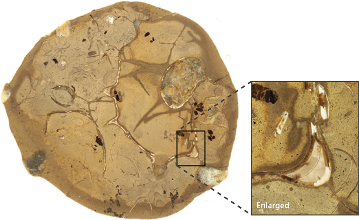
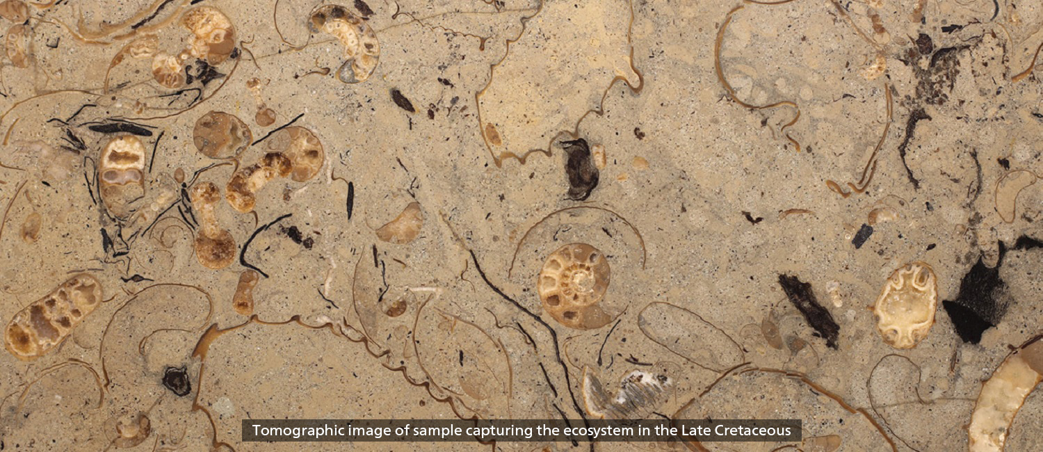
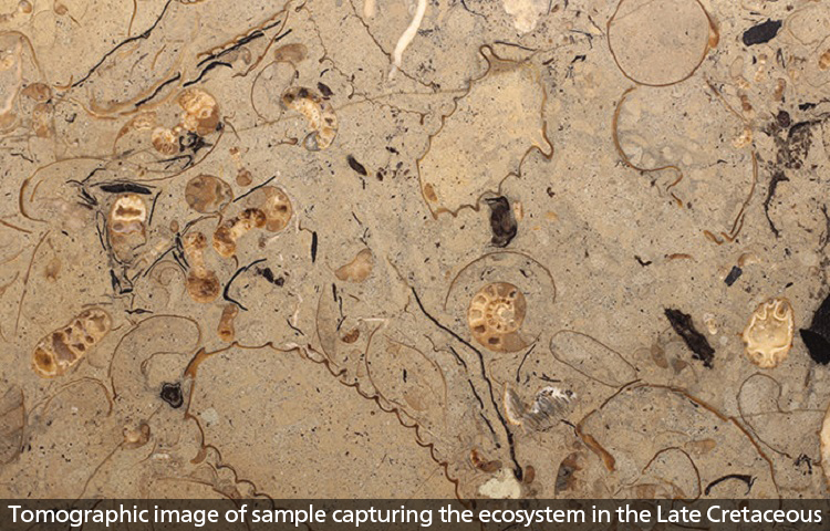
Tomographic image of sample captured with new tomographic technique
The image data obtained via this technique achieves significantly higher resolution than through previous methods. The amount of information contained in each voxel of a 3D model is about 100 million times that produced by a CT scanner.
With this newly-developed imaging technique, the probability of detecting fossils is expected to be much higher than conventional methods used thus far. In addition to paleontological researches, this cutting-edge technology also holds potential for use in a wide variety of fields and will help foster innovation in natural science research.
Canon cameras and lenses—capturing images of ancient life with superb accuracy
The process of polishing samples and photographing them might seem relatively straightforward. However, developing the equipment itself wasn’t so simple; stabilized image capture was a key requirement.
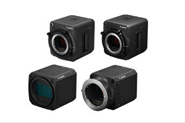
In order to produce a precise 3D model, it is necessary to maintain colors and brightness throughout the tomographic imaging process, where images are captured repeatedly for more than 1,000 times. Dr. Iba and his research team tested a variety of digital cameras and macro lenses, but were unable to obtain consistent images with most of the combinations until they tried out the Canon MP-E 65mm f/2.8 1-5x Macro Photo and EF 100mm f/2.8L Macro IS USM lenses, which, when combined with the EOS 5Ds, deliver consistent exposures and better stability than any other macro lenses tested thus far.
Equipped with a 35mm film-equivalent full-frame CMOS sensor with a resolution of approximately 50.6 megapixels, the Canon EOS 5Ds exceeds 8K (8688 x 5792 pixels), whereas the MP-E 65mm f/2.8 1-5x Macro Photo supports macro photography at up to 5x magnification. Used together, they deliver a level of resolving power surpassing that of stereomicroscopes.
Canon 8K displays deliver visuals just like what we see with the naked eye
8K displays are essential for observation and analysis of ultra-high-resolution images captured using next-generation 3D imaging techniques. These displays can displaying more than 16 times the information of full HD resolution, enabling the study of samples down to the microscopic structure. While conventional displays also allow users to examine details by magnifying the image, the 8K display is capable of subsampling—downscaling the data so as to enable full-screen display of the image when the resolution limit is exceeded.
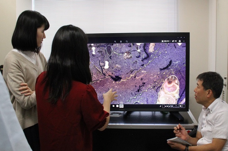
By rendering images in ultra-high-resolution that exceeds the resolving power of human eyes and employs a proprietary backlight control technology, Canon 8K monitors deliver images that accurately reproduce the dimensionality and texture of what we actually see. “Usually, we find new clues through on-site observation of the actual samples, but Canon’s 8K monitors deliver realistic results, as if we are looking at the actual object, which helps us make new discoveries in our research. It is fundamentally different from commercial-grade 8K displays,” says Dr. Iba. The remarkable color reproduction and contrast performance is able to meet the rigorous demand of professionals, allowing them to determine even the subtlest changes and finest structures.
A major driving force for cutting-edge innovations in natural science research
In addition to palaeontology, next-generation 3D imaging technology can also be employed in other fields such as medicine. Since information is digitally generated, findings and analyses can be shared in real time with other researchers around the world. Furthermore, by enabling researchers specializing in different areas to easily access high-quality information, we can also expect innovative breakthroughs made possible by interdisciplinary collaboration. Canon will continue to further enhance its 8K imaging technology and contribute to the progress of leading-edge research around the world.