
Visualizing functional diseases that were difficult to determine with conventional CT, and contributing to extending healthy lifespans
A CT system for scanning standing patients
A CT scanner uses X-rays to obtain cross-sectional images of the body, enabling visualization of lesions just a few millimeters in size within the body. Until now, examinations were generally performed with the patient lying on a couch, making it difficult to diagnose diseases that cause pain while standing, such as knee osteoarthritis or disc herniation. A new CT scanner that allows examinations of standing and seated patients is attracting interest.
June 27, 2024
Upright CT for early detection of decline in physical function in a super-aged society
Keio University Hospital is a designated Special Functioning Hospital that provides advanced medical care, develops and evaluates advanced medical technologies, and treats over one million patients per year. One of Japan's leading medical institutions, the hospital is also certified as a clinical research core hospital* that promotes clinical research and trials necessary for the development of innovative pharmaceuticals and medical devices originating in Japan.
Keio University Hospital is conducting clinical research in a variety of fields, including industry-academia collaboration, and one of these research themes is an upright CT scanner jointly developed by Professor Masahiro Jinzaki of the Department of Radiology, Keio University School of Medicine, and Canon Medical Systems.
The first clinical system was introduced at Keio University Hospital in 2017, with ongoing studies on its clinical usefulness. We spoke to Professor Jinzaki, system inventor and leader of the research group, about the background of upright CT.
- *Certified by the Ministry of Health, Labour and Welfare. As of the end of April 2023, 15 medical institutions have met the strict standards for certification.

Keio University Hospital

Professor, Department of Radiology, Keio University School of Medicine
Vice Director of Keio University Hospital
Masahiro Jinzaki
In the past, diagnostic imaging using CT was typically combined with contrast X-ray examinations. As CT technology evolved, 2D (single-slice) scanning was succeeded by 3D (volume) scanning and, in the late 2000s, 4D (dynamic) scanning. Once dynamic CT imaging became possible, contrast X-ray examinations were gradually replaced by CT exams.
"Generally, CT scanning is performed with the patient lying on a couch, but humans spend most of their time standing. Because the weight loading on the body differs between lying and standing, we thought there might be cases where accurate diagnosis couldn't be made with a conventional CT scan performed on a patient lying down. In addition, it isn't possible to obtain natural images of movements such as swallowing or walking in a scan performed on a patient lying down, and we felt that it was difficult to properly evaluate patients unless they were standing."
Around this time, Japan was becoming a super-aged society in which one in five people were over 65 years old, and terms such as "healthy lifespan" and "quality of life" were coming to public attention. As average lifespans increased, interest shifted from "living a long life without getting sick" to "living a long and healthy life." In the field of diagnostics, in addition to the early detection and diagnosis of "organic diseases" such as cancer and arteriosclerosis, where symptoms appear in the organs themselves, attention also shifted to "functional diseases," whose symptoms appear due to a decline in muscle strength and motor function caused by aging.
Providing quick and safe examinations for patients
Professor Jinzaki, who had been nurturing the idea of upright CT, proposed joint development to Canon Medical Systems (formerly Toshiba Medical Systems) in 2012, and after two years of consideration and preparation, the development project began in 2014. Although a prototype was soon completed, the road to the first clinical trial in 2017 was not smooth.

Professor Jinzaki experiencing the size of the system using a life-size cardboard model (photographed in 2015)
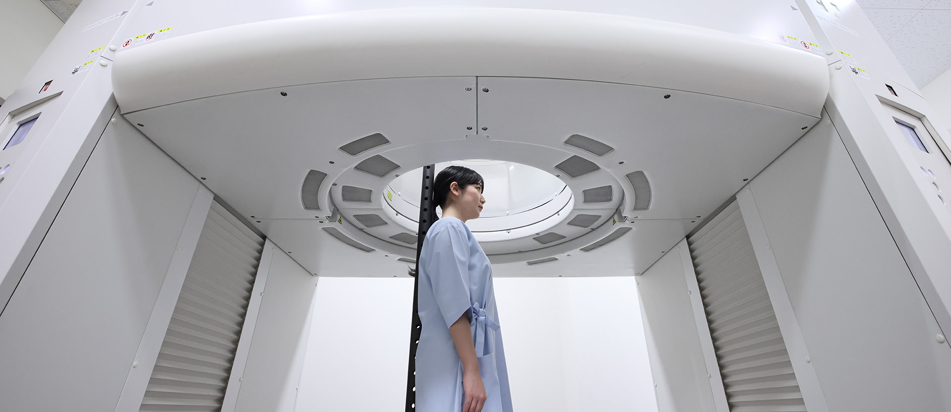
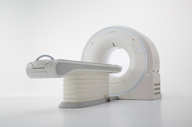
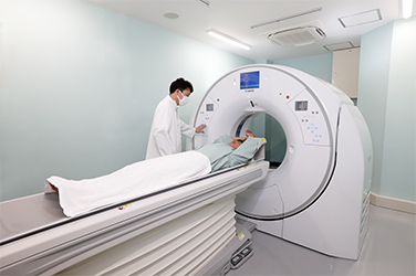
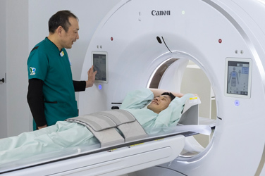
In conventional CT, the patient lies on a couch that is moved into a doughnut-shaped gantry for scanning. With upright CT, the patient stands in the center of a doughnut-shaped gantry or is seated in a special chair, and the gantry itself moves up and down during scanning.
"When developing upright CT, we focused on how to maintain the patient’s posture during scanning. The scan cannot be performed with the patient leaning against the system. An important focus in upright CT is how to keep the patient stable in a natural posture during the scan."
Even when people think they are standing straight, they may be swaying, and if scanning takes a long time, the image will be blurred. This is particularly likely to occur for elderly patients with declining motor function. Repeated testing of parts to support the patient was required until we finally discovered that lightly resting the back against a support pillar would provide stability.
Upright CT scan in which the patient places their back against a support pillar (Video, 17 seconds) *No audio
*In order to view videos, it is necessary to consent to the use of cookies by our website. If the videos are not displayed, please click the "Cookie Settings" and accept cookies.
Canon Medical Systems technology is utilized in the mechanism that moves the gantry with a mass of approximately two tons up and down at high speed while keeping it horizontal. Canon Medical Systems has developed the world's first* CT scanner that can capture a 16-cm wide image field in a single rotation around the patient. By adding new controls to this technology, a vertical-movement gantry for upright CT was made possible. With upright CT, it is now possible to scan the abdomen in approximately five seconds, ensuring both short examination times and safety.
"The concept of upright CT emerged in the late 1970s, but it was never implemented. In those days, scan times were long, and it would have been unrealistic to scan a standing patient. Even with the scan times possible around the year 2000, I think it would still have been difficult to achieve. When we wanted to create an upright CT scanner, the high-speed and high-definition CT technology that Canon Medical Systems already possessed was a great advantage."
- *As of January 2018 (according to research by Canon)
Expectations for identification of the causes of pain that can't be determined with conventional CT
"My patient was so happy when the cause of the pain was finally identified," said Professor Jinzaki, as he shared with us the words of the first patient.
The pain occurred only when the patient was standing. Examination with conventional systems had not been able to determine the cause of the pain, so this patient was finally referred to Keio University Hospital. An upright CT scan revealed the cause of the pain, and surgery was performed. Professor Jinzaki says he will never forget the joy he saw in the patient's face when he directly spoke with the patient after the operation.
Upright CT is expected to enable proper diagnosis, even for patients who have previously given up on investigating the cause of their symptoms, and help them to maintain and extend their healthy lifespan. Furthermore, upright CT is well received by patients, as the examination procedure is simpler than conventional CT. Previously, the positioning procedure required that a medical professional attend to the patient in the examination room, have them lie on a couch, and set them in an appropriate scan position. With upright CT, however, patients only need to stand in a designated position. There is no need for them to remove their shoes or lie down, and the examination time can be reduced by approximately 40%. For medical facilities, this will enable them to conduct 1.6 times as many examinations in the same amount of time, and it will also reduce contact between medical staff and patients, thereby reducing the risk of infection.
Enabling disease prevention with examination results and contributing to extending healthy lifespans
The usefulness of upright CT was demonstrated by Professor Jinzaki and his research group. In 2023, upright CT was also introduced to the Center for Preventive Medicine at Keio University and used for health checkups. Concerning the future of upright CT, Professor Jinzaki says, "In an aging society, it is important to detect functional decline at an early point in order to maintain health. For example, by tracking changes in bone and muscle mass over time, we can predict the risk of future decline and take measures to prevent it, or quantify posture and provide advice, and we hope to use the test results obtained by upright CT to help prevent disease and contribute to extending healthy lifespans."
Canon Medical Systems will continue to improve its technology in order to realize Professor Jinzaki's vision of the future of medical care.


